3IB2

structure of the complex of C-terminal half (C-lobe) of bovine lactoferrin with alpha-methyl-4-(2-methylpropyl) benzene acetic acid


Experimental Data Snapshot

  • Method: X-RAY DIFFRACTION
  • Resolution: 2.29 Å
  • R-Value Free: 0.220 
  • R-Value Work: 0.165 
  • R-Value Observed: 0.168 

wwPDB Validation   3D Report Full Report


Ligand Structure Quality Assessment 


This is version 2.1 of the entry. See complete history


Literature

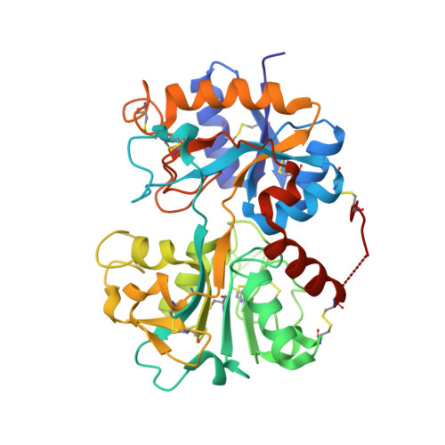
The structural basis for the prevention of nonsteroidal antiinflammatory drug-induced gastrointestinal tract damage by the C-lobe of bovine colostrum lactoferrin.

Mir, R.Singh, N.Vikram, G.Kumar, R.P.Sinha, M.Bhushan, A.Kaur, P.Srinivasan, A.Sharma, S.Singh, T.P.

(2009) Biophys J 97: 3178-3186

  • DOI: https://doi.org/10.1016/j.bpj.2009.09.030
  • Primary Citation of Related Structures:  
    3IAZ, 3IB0, 3IB1, 3IB2

  • PubMed Abstract: 

    Nonsteroidal antiinflammatory drugs (NSAIDs), due to their good efficacy in the treatment of pain, inflammation, and fever, are among the most prescribed class of medicines in the world. The main drawback of NSAIDs is that they induce gastric complications such as peptic ulceration and injury to the intestine. Four NSAIDs, indomethacin, diclofenac, aspirin, and ibuprofen were selected to induce gastropathy in mouse models. It was found that the addition of C-terminal half of bovine lactoferrin (C-lobe) reversed the NSAID-induced injuries to the extent of 47-70% whereas the coadministration of C-lobe prevented it significantly. The C-lobe was prepared proteolytically using serine proteases. The binding studies of C-lobe with NSAIDs showed that these compounds bind to C-lobe with affinities ranging from 2.6 to 4.8 x 10(-4) M. The complexes of C-lobe were prepared with the above four NSAIDs. All four complexes were crystallized and their detailed three-dimensional structures were determined using x-ray crystallographic method. The structures showed that all the four NSAID molecules bound to C-lobe at the newly identified ligand binding site in C-lobe that is formed involving two alpha-helices, alpha10 and alpha11. The ligand binding site is separated from the well known iron binding site by the longest and the most stable beta-strand, betaj, in the structure. Similar results were also obtained with the full length lactoferrin molecule. This novel, to our knowledge, binding site in C-lobe of lactoferrin shows a good complementarity for the acidic and lipophilic compounds such as NSAIDs. We believe this indicates that C-lobe of lactoferrin can be exploited for the prevention of NSAID-induced gastropathy.


  • Organizational Affiliation

    Department of Biophysics, All India Institute of Medical Sciences, New Delhi, India.


Macromolecules
Find similar proteins by:  (by identity cutoff)  |  3D Structure
Entity ID: 1
MoleculeChains Sequence LengthOrganismDetailsImage
Lactotransferrin345Bos taurusMutation(s): 0 
EC: 3.4.21
UniProt
Find proteins for P24627 (Bos taurus)
Explore P24627 
Go to UniProtKB:  P24627
Entity Groups  
Sequence Clusters30% Identity50% Identity70% Identity90% Identity95% Identity100% Identity
UniProt GroupP24627
Sequence Annotations
Expand
  • Reference Sequence
Oligosaccharides

Help

Entity ID: 2
MoleculeChains Length2D Diagram Glycosylation3D Interactions
alpha-D-mannopyranose-(1-4)-2-acetamido-2-deoxy-beta-D-glucopyranose-(1-4)-2-acetamido-2-deoxy-beta-D-glucopyranose
B
3N-Glycosylation
Glycosylation Resources
GlyTouCan:  G62182OO
GlyCosmos:  G62182OO
GlyGen:  G62182OO
Entity ID: 3
MoleculeChains Length2D Diagram Glycosylation3D Interactions
2-acetamido-2-deoxy-beta-D-glucopyranose-(1-4)-2-acetamido-2-deoxy-beta-D-glucopyranose
C, D
2N-Glycosylation
Glycosylation Resources
GlyTouCan:  G42666HT
GlyCosmos:  G42666HT
GlyGen:  G42666HT
Small Molecules
Ligands 5 Unique
IDChains Name / Formula / InChI Key2D Diagram3D Interactions
IBP
Query on IBP

Download Ideal Coordinates CCD File 
E [auth A]IBUPROFEN
C13 H18 O2
HEFNNWSXXWATRW-JTQLQIEISA-N
SO4
Query on SO4

Download Ideal Coordinates CCD File 
J [auth A]SULFATE ION
O4 S
QAOWNCQODCNURD-UHFFFAOYSA-L
ZN
Query on ZN

Download Ideal Coordinates CCD File 
H [auth A],
I [auth A]
ZINC ION
Zn
PTFCDOFLOPIGGS-UHFFFAOYSA-N
CO3
Query on CO3

Download Ideal Coordinates CCD File 
G [auth A]CARBONATE ION
C O3
BVKZGUZCCUSVTD-UHFFFAOYSA-L
FE
Query on FE

Download Ideal Coordinates CCD File 
F [auth A]FE (III) ION
Fe
VTLYFUHAOXGGBS-UHFFFAOYSA-N
Binding Affinity Annotations 
IDSourceBinding Affinity
IBP Binding MOAD:  3IB2 Kd: 4.80e+5 (nM) from 1 assay(s)
Experimental Data & Validation

Experimental Data

  • Method: X-RAY DIFFRACTION
  • Resolution: 2.29 Å
  • R-Value Free: 0.220 
  • R-Value Work: 0.165 
  • R-Value Observed: 0.168 
  • Space Group: P 1 21 1
Unit Cell:
Length ( Å )Angle ( ˚ )
a = 63.319α = 90
b = 50.48β = 107.75
c = 66.116γ = 90
Software Package:
Software NamePurpose
DENZOdata reduction
AMoREphasing
REFMACrefinement
AUTOMARdata reduction
SCALEPACKdata scaling

Structure Validation

View Full Validation Report


Ligand Structure Quality Assessment 


Entry History 

Deposition Data

Revision History  (Full details and data files)

  • Version 1.0: 2009-07-28
    Type: Initial release
  • Version 1.1: 2011-07-13
    Changes: Non-polymer description, Version format compliance
  • Version 2.0: 2020-07-29
    Type: Remediation
    Reason: Carbohydrate remediation
    Changes: Advisory, Atomic model, Data collection, Derived calculations, Structure summary
  • Version 2.1: 2023-11-01
    Changes: Data collection, Database references, Refinement description, Structure summary