3IGP

Structure of inhibitor binding to Carbonic Anhydrase II


Experimental Data Snapshot

  • Method: X-RAY DIFFRACTION
  • Resolution: 1.65 Å
  • R-Value Free: 0.196 
  • R-Value Work: 0.159 
  • R-Value Observed: 0.161 

wwPDB Validation   3D Report Full Report


Ligand Structure Quality Assessment 


This is version 1.3 of the entry. See complete history


Literature

Identification of 3,4-Dihydroisoquinoline-2(1H)-sulfonamides as Potent Carbonic Anhydrase Inhibitors: Synthesis, Biological Evaluation, and Enzyme-Ligand X-ray Studies.

Gitto, R.Agnello, S.Ferro, S.De Luca, L.Vullo, D.Brynda, J.Mader, P.Supuran, C.T.Chimirri, A.

(2010) J Med Chem 

  • DOI: https://doi.org/10.1021/jm9014026
  • Primary Citation of Related Structures:  
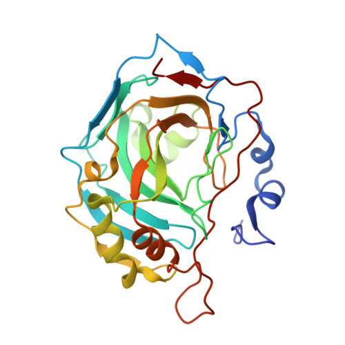
    3IGP

  • PubMed Abstract: 

    Following previous studies we herein report the exploration of the carbonic anhydrase (CA, EC 4.2.1.1) inhibitory effects and enzyme selectivity of a small class of 1-(cyclo)alkylisoquinolines containing a sulfonamide function considered a key feature for inhibiting CA. The results of enzymatic assays against human (h) CA isoforms, hCA I and hCA II (cytosolic, ubiquitous enzymes), hCA IX (transmembrane, tumor-associated), and hCA XIV (transmembrane), suggested that the presence of C-1 small substituents on isoquinoline scaffold controls both inhibitory potency and selectivity. Some derivatives showed potent hCA IX and hCA XIV inhibitory effects at nanomolar concentrations as well as low affinity for the ubiquitous hCA II. Moreover, we report the X-ray crystal structure of one of these derivatives in complex with dominant human isoform II, thus confirming the sulfonamide--zinc interactions. Finally, the results of docking experiments suggested the hypothetic interactions in the catalytic binding site for the most active and selective hCA IX and hCA XIV inhibitor.


  • Organizational Affiliation

    Dipartimento Farmaco-Chimico, Universita di Messina, Viale Annunziata, I-98168 Messina, Italy. rgitto@pharma.unime.it


Macromolecules
Find similar proteins by:  (by identity cutoff)  |  3D Structure
Entity ID: 1
MoleculeChains Sequence LengthOrganismDetailsImage
Carbonic anhydrase 2260Homo sapiensMutation(s): 0 
EC: 4.2.1.1
UniProt & NIH Common Fund Data Resources
Find proteins for P00918 (Homo sapiens)
Explore P00918 
Go to UniProtKB:  P00918
PHAROS:  P00918
GTEx:  ENSG00000104267 
Entity Groups  
Sequence Clusters30% Identity50% Identity70% Identity90% Identity95% Identity100% Identity
UniProt GroupP00918
Sequence Annotations
Expand
  • Reference Sequence
Small Molecules
Binding Affinity Annotations 
IDSourceBinding Affinity
DT7 PDBBind:  3IGP Ki: 94.5 (nM) from 1 assay(s)
Binding MOAD:  3IGP Ki: 32.8 (nM) from 1 assay(s)
Experimental Data & Validation

Experimental Data

  • Method: X-RAY DIFFRACTION
  • Resolution: 1.65 Å
  • R-Value Free: 0.196 
  • R-Value Work: 0.159 
  • R-Value Observed: 0.161 
  • Space Group: P 1 21 1
Unit Cell:
Length ( Å )Angle ( ˚ )
a = 42.31α = 90
b = 41.27β = 104.22
c = 72.04γ = 90
Software Package:
Software NamePurpose
MAR345data collection
MOLREPphasing
REFMACrefinement
MOSFLMdata reduction
SCALAdata scaling

Structure Validation

View Full Validation Report


Ligand Structure Quality Assessment 


Entry History 

Deposition Data

Revision History  (Full details and data files)

  • Version 1.0: 2010-03-09
    Type: Initial release
  • Version 1.1: 2011-07-13
    Changes: Version format compliance
  • Version 1.2: 2018-08-08
    Changes: Data collection
  • Version 1.3: 2023-11-01
    Changes: Data collection, Database references, Derived calculations, Refinement description