3MCO

Crystal Structure of the 6-hyroxymethyl-7,8-dihydropterin pyrophosphokinase dihydropteroate synthase bifunctional enzyme from Francisella tularensis


Experimental Data Snapshot

  • Method: X-RAY DIFFRACTION
  • Resolution: 2.30 Å
  • R-Value Free: 0.294 
  • R-Value Work: 0.241 
  • R-Value Observed: 0.243 

wwPDB Validation   3D Report Full Report


Ligand Structure Quality Assessment 


This is version 1.3 of the entry. See complete history


Literature

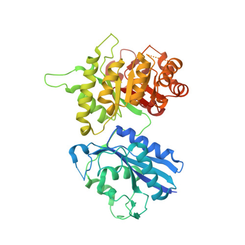
Crystal structure of the 6-hydroxymethyl-7,8-dihydropterin pyrophosphokinase.dihydropteroate synthase bifunctional enzyme from Francisella tularensis.

Pemble, C.W.Mehta, P.K.Mehra, S.Li, Z.Nourse, A.Lee, R.E.White, S.W.

(2010) PLoS One 5: e14165-e14165

  • DOI: https://doi.org/10.1371/journal.pone.0014165
  • Primary Citation of Related Structures:  
    3MCM, 3MCN, 3MCO

  • PubMed Abstract: 

    The 6-hydroxymethyl-7,8-dihydropterin pyrophosphokinase (HPPK) and dihydropteroate synthase (DHPS) enzymes catalyze sequential metabolic reactions in the folate biosynthetic pathway of bacteria and lower eukaryotes. Both enzymes represent validated targets for the development of novel anti-microbial therapies. We report herein that the genes which encode FtHPPK and FtDHPS from the biowarfare agent Francisella tularensis are fused into a single polypeptide. The potential of simultaneously targeting both modules with pterin binding inhibitors prompted us to characterize the molecular details of the multifunctional complex. Our high resolution crystallographic analyses reveal the structural organization between FtHPPK and FtDHPS which are tethered together by a short linker. Additional structural analyses of substrate complexes reveal that the active sites of each module are virtually indistinguishable from those of the monofunctional enzymes. The fused bifunctional enzyme therefore represents an excellent vehicle for finding inhibitors that engage the pterin binding pockets of both modules that have entirely different architectures. To demonstrate that this approach has the potential of producing novel two-hit inhibitors of the folate pathway, we identify and structurally characterize a fragment-like molecule that simultaneously engages both active sites. Our study provides a molecular framework to study the enzyme mechanisms of HPPK and DHPS, and to design novel and much needed therapeutic compounds to treat infectious diseases.


  • Organizational Affiliation

    Department of Structural Biology, St. Jude Children's Research Hospital, and Department of Molecular Sciences, University of Tennessee Health Science Center, Memphis, Tennessee, United States of America.


Macromolecules
Find similar proteins by:  (by identity cutoff)  |  3D Structure
Entity ID: 1
MoleculeChains Sequence LengthOrganismDetailsImage
2-amino-4-hydroxy-6-hydroxymethyldihydropteridine pyrophosphokinase/dihydropteroate synthase
A, B
442Francisella tularensis subsp. holarctica LVSMutation(s): 0 
Gene Names: folP/KFTL_1265
Entity Groups  
Sequence Clusters30% Identity50% Identity70% Identity90% Identity95% Identity100% Identity
Sequence Annotations
Expand
  • Reference Sequence
Small Molecules
Ligands 3 Unique
IDChains Name / Formula / InChI Key2D Diagram3D Interactions
APC
Query on APC

Download Ideal Coordinates CCD File 
E [auth A],
J [auth B]
DIPHOSPHOMETHYLPHOSPHONIC ACID ADENOSYL ESTER
C11 H18 N5 O12 P3
CAWZRIXWFRFUQB-IOSLPCCCSA-N
PH2
Query on PH2

Download Ideal Coordinates CCD File 
C [auth A],
D [auth A],
H [auth B],
I [auth B]
2-AMINO-6-HYDROXYMETHYL-7,8-DIHYDRO-3H-PTERIDIN-4-ONE
C7 H9 N5 O2
CQQNNQTXUGLUEV-UHFFFAOYSA-N
MG
Query on MG

Download Ideal Coordinates CCD File 
F [auth A],
G [auth A],
K [auth B],
L [auth B]
MAGNESIUM ION
Mg
JLVVSXFLKOJNIY-UHFFFAOYSA-N
Experimental Data & Validation

Experimental Data

  • Method: X-RAY DIFFRACTION
  • Resolution: 2.30 Å
  • R-Value Free: 0.294 
  • R-Value Work: 0.241 
  • R-Value Observed: 0.243 
  • Space Group: P 1
Unit Cell:
Length ( Å )Angle ( ˚ )
a = 42.521α = 82
b = 58.472β = 81
c = 109.327γ = 68.08
Software Package:
Software NamePurpose
DENZOdata reduction
SCALEPACKdata scaling
PHASERphasing
PHENIXrefinement
PDB_EXTRACTdata extraction

Structure Validation

View Full Validation Report


Ligand Structure Quality Assessment 


Entry History 

Deposition Data

Revision History  (Full details and data files)

  • Version 1.0: 2010-12-22
    Type: Initial release
  • Version 1.1: 2011-07-13
    Changes: Version format compliance
  • Version 1.2: 2017-11-08
    Changes: Refinement description
  • Version 1.3: 2023-09-06
    Changes: Data collection, Database references, Derived calculations, Refinement description