3MGX

Crystal Structure of P450 OxyD that is involved in the Biosynthesis of Vancomycin-type Antibiotics


Experimental Data Snapshot

  • Method: X-RAY DIFFRACTION
  • Resolution: 2.10 Å
  • R-Value Free: 0.233 
  • R-Value Work: 0.208 
  • R-Value Observed: 0.209 

wwPDB Validation   3D Report Full Report


Ligand Structure Quality Assessment 


This is version 1.2 of the entry. See complete history


Literature

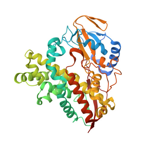
Structural characterization of oxyd, a cytochrome p450 involved in {beta}-hydroxytyrosine formation in vancomycin biosynthesis

Cryle, M.J.Meinhart, A.Schlichting, I.

(2010) J Biol Chem 285: 24562-24574

  • DOI: https://doi.org/10.1074/jbc.M110.131904
  • Primary Citation of Related Structures:  
    3MGX

  • PubMed Abstract: 

    The cytochrome P450 OxyD from the balhimycin glycopeptide antibiotic biosynthetic operon of Amycolatopsis mediterranei is involved in the biosynthesis of the modified amino acid beta-R-hydroxytyrosine, an essential precursor for biosynthesis of the vancomycin-type aglycone. OxyD binds the substrate tyrosine not free in solution, but rather covalently linked to the carrier protein (CP) domain of the non-ribosomal peptide synthase BpsD, exhibiting micromolar binding affinity to a tyrosine-loaded carrier protein construct. The crystal structure of OxyD was determined to 2.1-A resolution, revealing a potential binding site for the carrier protein-bound substrate in a different orientation to that seen with the acyl carrier protein-bound P450(BioI) (Cryle, M. J., and Schlichting, I. (2008) Proc. Natl. Acad. Sci. U.S.A. 105, 15696-15701). A series of residues were identified across known aminoacyl-CP-oxidizing P450s that are highly conserved and cluster in the active site or potential CP binding site of OxyD. These residues appear to be characteristic for aminoacyl-CP-oxidizing P450s, allowing sequence based identification of P450 function for this subgroup of P450s that play vital roles in the biosyntheses of many important natural products in addition to the vancomycin-type antibiotics. The ability to analyze such P450 function based upon sequence data alone should prove an important tool in the analysis and identification of new medicinally relevant biomolecules.


  • Organizational Affiliation

    Department of Biomolecular Mechanisms, Max-Planck Institute for Medical Research, Jahnstrasse 29, 69120 Heidelberg, Germany. Max.Cryle@mpimf-heidelberg.mpg.de


Macromolecules
Find similar proteins by:  (by identity cutoff)  |  3D Structure
Entity ID: 1
MoleculeChains Sequence LengthOrganismDetailsImage
Putative P450 monooxygenase
A, B
415Amycolatopsis balhimycinaMutation(s): 0 
Gene Names: oxyD
EC: 1.14.14.1
UniProt
Find proteins for Q939Y1 (Amycolatopsis balhimycina)
Explore Q939Y1 
Go to UniProtKB:  Q939Y1
Entity Groups  
Sequence Clusters30% Identity50% Identity70% Identity90% Identity95% Identity100% Identity
UniProt GroupQ939Y1
Sequence Annotations
Expand
  • Reference Sequence
Small Molecules
Ligands 2 Unique
IDChains Name / Formula / InChI Key2D Diagram3D Interactions
HEM
Query on HEM

Download Ideal Coordinates CCD File 
C [auth A],
I [auth B]
PROTOPORPHYRIN IX CONTAINING FE
C34 H32 Fe N4 O4
KABFMIBPWCXCRK-RGGAHWMASA-L
GOL
Query on GOL

Download Ideal Coordinates CCD File 
D [auth A]
E [auth A]
F [auth A]
G [auth A]
H [auth A]
D [auth A],
E [auth A],
F [auth A],
G [auth A],
H [auth A],
J [auth B],
K [auth B],
L [auth B],
M [auth B],
N [auth B]
GLYCEROL
C3 H8 O3
PEDCQBHIVMGVHV-UHFFFAOYSA-N
Experimental Data & Validation

Experimental Data

  • Method: X-RAY DIFFRACTION
  • Resolution: 2.10 Å
  • R-Value Free: 0.233 
  • R-Value Work: 0.208 
  • R-Value Observed: 0.209 
  • Space Group: P 1 21 1
Unit Cell:
Length ( Å )Angle ( ˚ )
a = 65.85α = 90
b = 61.05β = 102.48
c = 100.64γ = 90
Software Package:
Software NamePurpose
XDSdata scaling
PHASERphasing
REFMACrefinement
XDSdata reduction
XSCALEdata scaling

Structure Validation

View Full Validation Report


Ligand Structure Quality Assessment 


Entry History 

Deposition Data

Revision History  (Full details and data files)

  • Version 1.0: 2010-06-02
    Type: Initial release
  • Version 1.1: 2011-07-13
    Changes: Version format compliance
  • Version 1.2: 2023-11-01
    Changes: Data collection, Database references, Derived calculations, Refinement description