3OCZ

Structure of Recombinant Haemophilus influenzae e(P4) Acid Phosphatase Complexed with the inhibitor adenosine 5-O-thiomonophosphate


Experimental Data Snapshot

  • Method: X-RAY DIFFRACTION
  • Resolution: 1.35 Å
  • R-Value Free: 0.159 
  • R-Value Work: 0.138 
  • R-Value Observed: 0.139 

wwPDB Validation   3D Report Full Report


Ligand Structure Quality Assessment 


This is version 1.4 of the entry. See complete history


Literature

Structural basis of the inhibition of class C acid phosphatases by adenosine 5'-phosphorothioate.

Singh, H.Reilly, T.J.Tanner, J.J.

(2011) FEBS J 278: 4374-4381

  • DOI: https://doi.org/10.1111/j.1742-4658.2011.08360.x
  • Primary Citation of Related Structures:  
    3OCZ, 3SF0

  • PubMed Abstract: 

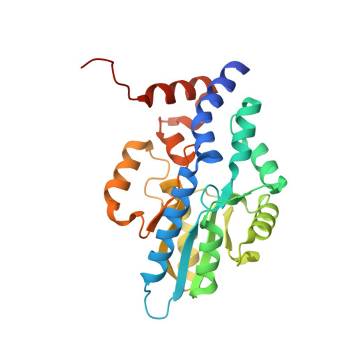
    The inhibition of phosphatases by adenosine 5'-phosphorothioate (AMPS) was first reported in the late 1960s; however, the structural basis for the inhibition has remained unknown. Here, it is shown that AMPS is a submicromolar inhibitor of class C acid phosphatases, a group of bacterial outer membrane enzymes belonging to the haloacid dehalogenase structural superfamily. Furthermore, the 1.35-Å resolution crystal structure of the inhibited recombinant Haemophilus influenzae class C acid phosphatase was determined; this is the first structure of a phosphatase complexed with AMPS. The conformation of AMPS is identical to that of the substrate 5'-AMP, except that steric factors force a rotation of the thiophosphoryl out of the normal phosphoryl-binding pocket. This conformation is catalytically nonproductive, because the P atom is not positioned optimally for nucleophilic attack by Asp64, and the O atom of the scissile O-P bond is too far from the Asp (Asp66) that protonates the leaving group. The structure of 5'-AMP complexed with the Asp64→Asn mutant enzyme was also determined at 1.35-Å resolution. This mutation induces the substrate to adopt the same nonproductive binding mode that is observed in the AMPS complex. In this case, electrostatic considerations, rather than steric factors, underlie the movement of the phosphoryl. The structures not only provide an explanation for the inhibition by AMPS, but also highlight the precise steric and electrostatic requirements of phosphoryl recognition by class C acid phosphatases. Moreover, the structure of the Asp64→Asn mutant illustrates how a seemingly innocuous mutation can cause an unexpected structural change.


  • Organizational Affiliation

    Department of Chemistry, University of Missouri-Columbia, Columbia, MO 65211, USA.


Macromolecules
Find similar proteins by:  (by identity cutoff)  |  3D Structure
Entity ID: 1
MoleculeChains Sequence LengthOrganismDetailsImage
Lipoprotein E262Haemophilus influenzaeMutation(s): 0 
Gene Names: helHI_0693ompP4
EC: 3.1.3.2
UniProt
Find proteins for P26093 (Haemophilus influenzae (strain ATCC 51907 / DSM 11121 / KW20 / Rd))
Explore P26093 
Go to UniProtKB:  P26093
Entity Groups  
Sequence Clusters30% Identity50% Identity70% Identity90% Identity95% Identity100% Identity
UniProt GroupP26093
Sequence Annotations
Expand
  • Reference Sequence
Small Molecules
Ligands 2 Unique
IDChains Name / Formula / InChI Key2D Diagram3D Interactions
SRA
Query on SRA

Download Ideal Coordinates CCD File 
C [auth A]ADENOSINE -5'-THIO-MONOPHOSPHATE
C10 H14 N5 O6 P S
UBCPYVAQZGCDJO-KQYNXXCUSA-N
MG
Query on MG

Download Ideal Coordinates CCD File 
B [auth A]MAGNESIUM ION
Mg
JLVVSXFLKOJNIY-UHFFFAOYSA-N
Binding Affinity Annotations 
IDSourceBinding Affinity
SRA Binding MOAD:  3OCZ Ki: 35 (nM) from 1 assay(s)
PDBBind:  3OCZ Ki: 35 (nM) from 1 assay(s)
Experimental Data & Validation

Experimental Data

  • Method: X-RAY DIFFRACTION
  • Resolution: 1.35 Å
  • R-Value Free: 0.159 
  • R-Value Work: 0.138 
  • R-Value Observed: 0.139 
  • Space Group: P 65 2 2
Unit Cell:
Length ( Å )Angle ( ˚ )
a = 98.179α = 90
b = 98.179β = 90
c = 107.02γ = 120
Software Package:
Software NamePurpose
DENZOdata reduction
SCALEPACKdata scaling
PHENIXrefinement
PDB_EXTRACTdata extraction
HKL-2000data reduction
HKL-2000data scaling
PHASERphasing

Structure Validation

View Full Validation Report


Ligand Structure Quality Assessment 


Entry History 

Deposition Data

Revision History  (Full details and data files)

  • Version 1.0: 2011-07-20
    Type: Initial release
  • Version 1.1: 2011-09-28
    Changes: Database references
  • Version 1.2: 2012-01-11
    Changes: Database references
  • Version 1.3: 2017-11-08
    Changes: Advisory, Refinement description
  • Version 1.4: 2023-09-06
    Changes: Advisory, Data collection, Database references, Derived calculations, Refinement description