3ONB

Bond breakage and relocation of a covalently bound bromine of IDD594 in a complex with hAR T113A mutant after extensive radiation dose


Experimental Data Snapshot

  • Method: X-RAY DIFFRACTION
  • Resolution: 1.45 Å
  • R-Value Free: 0.175 
  • R-Value Work: 0.117 
  • R-Value Observed: 0.117 

wwPDB Validation   3D Report Full Report


Ligand Structure Quality Assessment 


This is version 1.2 of the entry. See complete history


Literature

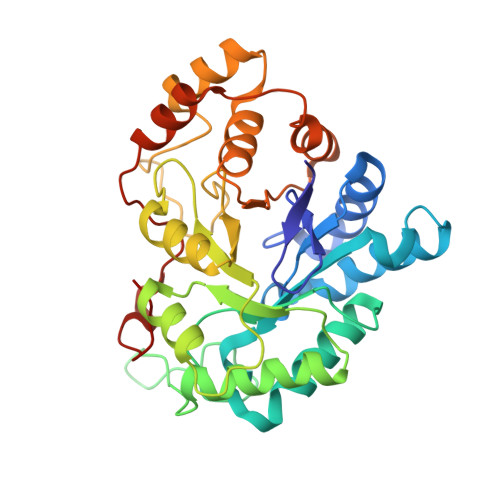
Radiation damage reveals promising interaction position

Koch, C.Heine, A.Klebe, G.

(2011) J Synchrotron Radiat 18: 782-789

  • DOI: https://doi.org/10.1107/S0909049511027920
  • Primary Citation of Related Structures:  
    3ONB, 3ONC

  • PubMed Abstract: 

    High-resolution structural data of protein inhibitor complexes are the key to rational drug design. Synchrotron radiation allows for atomic resolutions but is frequently accompanied by radiation damage to protein complexes. In this study a human aldose reductase mutant complexed with a bromine-substituted inhibitor was determined to atomic resolution [Protein Data Bank (PDB) code 3onc]. Though the radiation dose was moderate, a selective disruption of a bromine-inhibitor bond during the experiment was observed while the protein appears unaffected. A covalent bond to bromine is cleaved and the displaced atom is not scattered throughout the crystal but can most likely be assigned as a bromide to an additional difference electron density peak observed in the structure. The bromide relocates to an adjacent unoccupied site where promising interactions to protein residues stabilize its position. These findings were verified by a second similar structure determined with considerably higher radiation dose (PDB code 3onb).


  • Organizational Affiliation

    Philipps-Universität, Marburg, Germany.


Macromolecules
Find similar proteins by:  (by identity cutoff)  |  3D Structure
Entity ID: 1
MoleculeChains Sequence LengthOrganismDetailsImage
Aldose reductase315Homo sapiensMutation(s): 1 
Gene Names: alr2
EC: 1.1.1.21
UniProt & NIH Common Fund Data Resources
Find proteins for P15121 (Homo sapiens)
Explore P15121 
Go to UniProtKB:  P15121
PHAROS:  P15121
GTEx:  ENSG00000085662 
Entity Groups  
Sequence Clusters30% Identity50% Identity70% Identity90% Identity95% Identity100% Identity
UniProt GroupP15121
Sequence Annotations
Expand
  • Reference Sequence
Small Molecules
Binding Affinity Annotations 
IDSourceBinding Affinity
LDT BindingDB:  3ONB IC50: min: 3, max: 30 (nM) from 2 assay(s)
-TΔS: min: -2.26e+1, max: 7.99 (kJ/mol) from 3 assay(s)
ΔH: -4.62e+1 (kJ/mol) from 1 assay(s)
ΔG: min: -4.09e+1, max: -3.13e+1 (kJ/mol) from 4 assay(s)
Experimental Data & Validation

Experimental Data

  • Method: X-RAY DIFFRACTION
  • Resolution: 1.45 Å
  • R-Value Free: 0.175 
  • R-Value Work: 0.117 
  • R-Value Observed: 0.117 
  • Space Group: P 1 21 1
Unit Cell:
Length ( Å )Angle ( ˚ )
a = 49.08α = 90
b = 66.51β = 92.39
c = 47.14γ = 90
Software Package:
Software NamePurpose
MAR345dtbdata collection
SHELXmodel building
SHELXL-97refinement
HKL-2000data reduction
HKL-2000data scaling
SHELXphasing

Structure Validation

View Full Validation Report


Ligand Structure Quality Assessment 


Entry History 

Deposition Data

Revision History  (Full details and data files)

  • Version 1.0: 2011-08-17
    Type: Initial release
  • Version 1.1: 2011-11-02
    Changes: Database references
  • Version 1.2: 2024-03-20
    Changes: Data collection, Database references, Derived calculations