3OVQ

Crystal Structure of hRPE and D-Ribulose-5-Phospate Complex


Experimental Data Snapshot

  • Method: X-RAY DIFFRACTION
  • Resolution: 2.00 Å
  • R-Value Free: 0.191 
  • R-Value Work: 0.147 
  • R-Value Observed: 0.149 

wwPDB Validation   3D Report Full Report


Ligand Structure Quality Assessment 


This is version 1.3 of the entry. See complete history


Literature

Conversion of D-ribulose 5-phosphate to D-xylulose 5-phosphate: new insights from structural and biochemical studies on human RPE

Liang, W.G.Ouyang, S.Y.Shaw, N.Joachimiak, A.Zhang, R.G.Liu, Z.J.

(2011) FASEB J 25: 497-504

  • DOI: https://doi.org/10.1096/fj.10-171207
  • Primary Citation of Related Structures:  
    3OVP, 3OVQ, 3OVR

  • PubMed Abstract: 

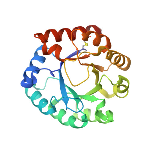
    The pentose phosphate pathway (PPP) confers protection against oxidative stress by supplying NADPH necessary for the regeneration of glutathione, which detoxifies H(2)O(2) into H(2)O and O(2). RPE functions in the PPP, catalyzing the reversible conversion of D-ribulose 5-phosphate to D-xylulose 5-phosphate and is an important enzyme for cellular response against oxidative stress. Here, using structural, biochemical, and functional studies, we show that human D-ribulose 5-phosphate 3-epimerase (hRPE) uses Fe(2+) for catalysis. Structures of the binary complexes of hRPE with D-ribulose 5-phosphate and D-xylulose 5-phosphate provide the first detailed molecular insights into the binding mode of physiological ligands and reveal an octahedrally coordinated Fe(2+) ion buried deep inside the active site. Human RPE folds into a typical (β/α)(8) triosephosphate isomerase (TIM) barrel with a loop regulating access to the active site. Two aspartic acids are well positioned to carry out the proton transfers in an acid-base type of reaction mechanism. Interestingly, mutating Ser-10 to alanine almost abolished the enzymatic activity, while L12A and M72A mutations resulted in an almost 50% decrease in the activity. The binary complexes of hRPE reported here will aid in the design of small molecules for modulating the activity of the enzyme and altering flux through the PPP.


  • Organizational Affiliation

    National Laboratory of Biomacromolecules, Institute of Biophysics, Chinese Academy of Sciences, Beijing 100101, China.


Macromolecules
Find similar proteins by:  (by identity cutoff)  |  3D Structure
Entity ID: 1
MoleculeChains Sequence LengthOrganismDetailsImage
Ribulose-phosphate 3-epimerase
A, B
228Homo sapiensMutation(s): 0 
Gene Names: RPEHUSSY-17
EC: 5.1.3.1
UniProt & NIH Common Fund Data Resources
Find proteins for Q96AT9 (Homo sapiens)
Explore Q96AT9 
Go to UniProtKB:  Q96AT9
PHAROS:  Q96AT9
GTEx:  ENSG00000197713 
Entity Groups  
Sequence Clusters30% Identity50% Identity70% Identity90% Identity95% Identity100% Identity
UniProt GroupQ96AT9
Sequence Annotations
Expand
  • Reference Sequence
Small Molecules
Ligands 3 Unique
IDChains Name / Formula / InChI Key2D Diagram3D Interactions
XPE
Query on XPE

Download Ideal Coordinates CCD File 
E [auth A]
F [auth A]
G [auth A]
H [auth A]
K [auth B]
E [auth A],
F [auth A],
G [auth A],
H [auth A],
K [auth B],
L [auth B],
M [auth B]
3,6,9,12,15,18,21,24,27-NONAOXANONACOSANE-1,29-DIOL
C20 H42 O11
DTPCFIHYWYONMD-UHFFFAOYSA-N
5RP
Query on 5RP

Download Ideal Coordinates CCD File 
D [auth A],
J [auth B]
RIBULOSE-5-PHOSPHATE
C5 H11 O8 P
FNZLKVNUWIIPSJ-UHNVWZDZSA-N
FE2
Query on FE2

Download Ideal Coordinates CCD File 
C [auth A],
I [auth B]
FE (II) ION
Fe
CWYNVVGOOAEACU-UHFFFAOYSA-N
Experimental Data & Validation

Experimental Data

  • Method: X-RAY DIFFRACTION
  • Resolution: 2.00 Å
  • R-Value Free: 0.191 
  • R-Value Work: 0.147 
  • R-Value Observed: 0.149 
  • Space Group: P 1 21 1
Unit Cell:
Length ( Å )Angle ( ˚ )
a = 61.785α = 90
b = 43.535β = 94.88
c = 72.269γ = 90
Software Package:
Software NamePurpose
PHASESphasing
PHENIXrefinement
HKL-2000data reduction
HKL-2000data scaling

Structure Validation

View Full Validation Report


Ligand Structure Quality Assessment 


Entry History 

Deposition Data

Revision History  (Full details and data files)

  • Version 1.0: 2011-03-09
    Type: Initial release
  • Version 1.1: 2011-07-13
    Changes: Version format compliance
  • Version 1.2: 2016-12-21
    Changes: Structure summary
  • Version 1.3: 2023-11-01
    Changes: Data collection, Database references, Derived calculations, Refinement description