A single acidic residue can guide binding site selection but does not govern QacR cationic-drug affinity.
Peters, K.M., Brooks, B.E., Schumacher, M.A., Skurray, R.A., Brennan, R.G., Brown, M.H.(2011) PLoS One 6: e15974-e15974
- PubMed: 21264225
- DOI: https://doi.org/10.1371/journal.pone.0015974
- Primary Citation of Related Structures:
3PM1 - PubMed Abstract:
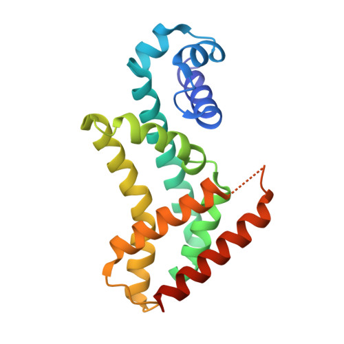
Structures of the multidrug-binding repressor protein QacR with monovalent and bivalent cationic drugs revealed that the carboxylate side-chains of E90 and E120 were proximal to the positively charged nitrogens of the ligands ethidium, malachite green and rhodamine 6G, and therefore may contribute to drug neutralization and binding affinity. Here, we report structural, biochemical and in vivo effects of substituting these glutamate residues. Unexpectedly, substitutions had little impact on ligand affinity or in vivo induction capabilities. Structures of QacR(E90Q) and QacR(E120Q) with ethidium or malachite green took similar global conformations that differed significantly from all previously described QacR-drug complexes but still prohibited binding to cognate DNA. Strikingly, the QacR(E90Q)-rhodamine 6G complex revealed two mutually exclusive rhodamine 6G binding sites. Despite multiple structural changes, all drug binding was essentially isoenergetic. Thus, these data strongly suggest that rather than contributing significantly to ligand binding affinity, the role of acidic residues lining the QacR multidrug-binding pocket is primarily to attract and guide cationic drugs to the "best available" positions within the pocket that elicit QacR induction.
Organizational Affiliation:
School of Biological Sciences, University of Sydney, Sydney, New South Wales, Australia.