pH effects on binding between the anthrax protective antigen and the host cellular receptor CMG2.
Rajapaksha, M., Lovell, S., Janowiak, B.E., Andra, K.K., Battaile, K.P., Bann, J.G.(2012) Protein Sci 21: 1467-1480
- PubMed: 22855243
- DOI: https://doi.org/10.1002/pro.2136
- Primary Citation of Related Structures:
3Q8A, 3Q8B, 3Q8C, 3Q8E, 3Q8F - PubMed Abstract:
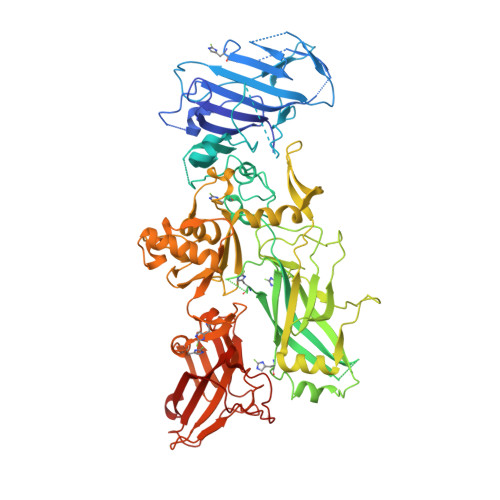
The anthrax protective antigen (PA) binds to the host cellular receptor capillary morphogenesis protein 2 (CMG2) with high affinity. To gain a better understanding of how pH may affect binding to the receptor, we have investigated the kinetics of binding as a function of pH to the full-length monomeric PA and to two variants: a 2-fluorohistidine-labeled PA (2-FHisPA), which is ∼1 pH unit more stable to variations in pH than WT, and an ∼1 pH unit less stable variant in which Trp346 in the domain 2β(3) -2β(4) loop is substituted with a Phe (W346F). We show using stopped-flow fluorescence that the binding rate increases as the pH is lowered for all proteins, with little influence on the rate of dissociation. In addition, we have crystallized PA and the two variants and examine the influence of pH on structure. In contrast to previous X-ray studies, the domain 2β(3) -2β(4) loop undergoes little change in structure from pH ∼8 to 5.5 for the WT protein, but for the 2-FHis labeled and W346F mutant there are changes in structure consistent with previous X-ray studies. In accord with pH stability studies, we find that the average B-factor values increase by ∼20-30% for all three proteins at low pH. Our results suggest that for the full-length PA, low pH increases the binding affinity, likely through a change in structure that favors a more "bound-like" conformation.
Organizational Affiliation:
Department of Chemistry, Wichita State University, Wichita, Kansas 67260-0051, USA.


