3QFU

Crystal structure of Yeast Hsp70 (Bip/kar2) complexed with ADP


Experimental Data Snapshot

  • Method: X-RAY DIFFRACTION
  • Resolution: 1.80 Å
  • R-Value Free: 0.238 
  • R-Value Work: 0.217 
  • R-Value Observed: 0.218 

wwPDB Validation   3D Report Full Report


Ligand Structure Quality Assessment 


This is version 1.3 of the entry. See complete history


Literature

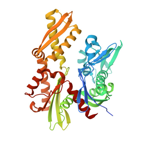
Structural analysis of the Sil1-Bip complex reveals the mechanism for Sil1 to function as a nucleotide-exchange factor.

Yan, M.Li, J.Sha, B.

(2011) Biochem J 438: 447-455

  • DOI: https://doi.org/10.1042/BJ20110500
  • Primary Citation of Related Structures:  
    3QFP, 3QFU, 3QML

  • PubMed Abstract: 

    Sil1 functions as a NEF (nucleotide-exchange factor) for the ER (endoplasmic reticulum) Hsp70 (heat-shock protein of 70 kDa) Bip in eukaryotic cells. Sil1 may catalyse the ADP release from Bip by interacting directly with the ATPase domain of Bip. In the present study we show the complex crystal structure of the yeast Bip and the NEF Sil1 at the resolution of 2.3 Å (1 Å=0.1 nm). In the Sil1-Bip complex structure, the Sil1 molecule acts as a 'clamp' which binds lobe IIb of the Bip ATPase domain. The binding of Sil1 causes the rotation of lobe IIb ~ 13.5° away from the ADP-binding pocket. The complex formation also induces lobe Ib to swing in the opposite direction by ~ 3.7°. These conformational changes open up the nucleotide-binding pocket in the Bip ATPase domain and disrupt the hydrogen bonds between Bip and bound ADP, which may catalyse ADP release. Mutation of the Sil1 residues involved in binding the Bip ATPase domain compromise the binding affinity of Sil1 to Bip, and these Sil1 mutants also abolish the ability to stimulate the ATPase activity of Bip.


  • Organizational Affiliation

    Department of Cell Biology, University of Alabama at Birmingham, Birmingham, AL 35294-0005, USA.


Macromolecules
Find similar proteins by:  (by identity cutoff)  |  3D Structure
Entity ID: 1
MoleculeChains Sequence LengthOrganismDetailsImage
78 kDa glucose-regulated protein homolog394Saccharomyces cerevisiaeMutation(s): 0 
Gene Names: KAR2GRP78SSD1YJL034WJ1248
UniProt
Find proteins for P16474 (Saccharomyces cerevisiae (strain ATCC 204508 / S288c))
Explore P16474 
Go to UniProtKB:  P16474
Entity Groups  
Sequence Clusters30% Identity50% Identity70% Identity90% Identity95% Identity100% Identity
UniProt GroupP16474
Sequence Annotations
Expand
  • Reference Sequence
Experimental Data & Validation

Experimental Data

  • Method: X-RAY DIFFRACTION
  • Resolution: 1.80 Å
  • R-Value Free: 0.238 
  • R-Value Work: 0.217 
  • R-Value Observed: 0.218 
  • Space Group: P 21 21 21
Unit Cell:
Length ( Å )Angle ( ˚ )
a = 63.367α = 90
b = 85.667β = 90
c = 94.921γ = 90
Software Package:
Software NamePurpose
HKL-2000data collection
PHASERphasing
REFMACrefinement
HKL-2000data reduction
HKL-2000data scaling

Structure Validation

View Full Validation Report


Ligand Structure Quality Assessment 


Entry History 

Deposition Data

Revision History  (Full details and data files)

  • Version 1.0: 2011-06-29
    Type: Initial release
  • Version 1.1: 2011-07-13
    Changes: Version format compliance
  • Version 1.2: 2011-09-07
    Changes: Database references
  • Version 1.3: 2023-09-13
    Changes: Data collection, Database references, Derived calculations, Refinement description