3TDT

COMPLEX OF TETRAHYDRODIPICOLINATE N-SUCCINYLTRANSFERASE WITH 2-AMINO-6-OXOPIMELATE AND COENZYME A


Experimental Data Snapshot

  • Method: X-RAY DIFFRACTION
  • Resolution: 2.00 Å
  • R-Value Free: 0.220 
  • R-Value Work: 0.152 
  • R-Value Observed: 0.154 

wwPDB Validation   3D Report Full Report


This is version 1.3 of the entry. See complete history


Literature

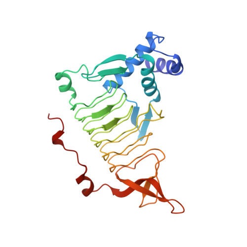
The conformational change and active site structure of tetrahydrodipicolinate N-succinyltransferase.

Beaman, T.W.Blanchard, J.S.Roderick, S.L.

(1998) Biochemistry 37: 10363-10369

  • DOI: https://doi.org/10.1021/bi980759b
  • Primary Citation of Related Structures:  
    2TDT, 3TDT

  • PubMed Abstract: 

    Tetrahydrodipicolinate (THDP) N-succinyltransferase catalyzes the conversion of tetrahydrodipicolinate and succinyl-CoA to L-2-(succinylamino)-6-oxopimelate and CoA. This reaction represents the committed step of the succinylase branch of the diaminopimelate/L-lysine biosynthetic pathway by which many bacteria synthesize meso-diaminopimelate, a component of peptidoglycan, and L-lysine from L-aspartate. The crystal structures of THDP succinyltransferase in complex with the substrate/cofactor pairs L-2-aminopimelate/coenzyme A and L-2-amino-6-oxopimelate/coenzyme A have been determined and refined to 2.0 A resolution. The active site of the enzyme is a long narrow groove located at the interface between two left-handed parallel beta-helix (LbetaH) structural domains of the trimeric enzyme. On binding the amino acid acceptor and cofactor, this groove is covered by residues from the C-terminus of one subunit and a flexible loop excluded from the LbetaH domain of an adjacent subunit to form a tunnel. This conformational change is directly related to interactions between the enzyme and the bound amino acid substrate and cofactor and serves to shield the ligands from bulk solvent and to orient the nucleophilic amino group of the amino acid acceptor toward the mercaptoethylamine group of the cofactor.


  • Organizational Affiliation

    Department of Biochemistry, Albert Einstein College of Medicine, Bronx, New York 10461, USA.


Macromolecules
Find similar proteins by:  (by identity cutoff)  |  3D Structure
Entity ID: 1
MoleculeChains Sequence LengthOrganismDetailsImage
TETRAHYDRODIPICOLINATE N-SUCCINYLTRANSFERASE274Mycobacterium tuberculosis variant bovisMutation(s): 0 
Gene Names: DAPD
EC: 2.3.1.117
UniProt
Find proteins for P56220 (Unknown prokaryotic organism)
Explore P56220 
Go to UniProtKB:  P56220
Entity Groups  
Sequence Clusters30% Identity50% Identity70% Identity90% Identity95% Identity100% Identity
UniProt GroupP56220
Sequence Annotations
Expand
  • Reference Sequence
Small Molecules
Ligands 2 Unique
IDChains Name / Formula / InChI Key2D Diagram3D Interactions
COA
Query on COA

Download Ideal Coordinates CCD File 
B [auth A]COENZYME A
C21 H36 N7 O16 P3 S
RGJOEKWQDUBAIZ-IBOSZNHHSA-N
26P
Query on 26P

Download Ideal Coordinates CCD File 
C [auth A]2-AMINO-6-OXOPIMELIC ACID
C7 H11 N O5
UKCSFKLWNHUBDY-BYPYZUCNSA-N
Experimental Data & Validation

Experimental Data

  • Method: X-RAY DIFFRACTION
  • Resolution: 2.00 Å
  • R-Value Free: 0.220 
  • R-Value Work: 0.152 
  • R-Value Observed: 0.154 
  • Space Group: H 3
Unit Cell:
Length ( Å )Angle ( ˚ )
a = 96α = 90
b = 96β = 90
c = 72.3γ = 120
Software Package:
Software NamePurpose
X-PLORmodel building
TNTrefinement
X-PLORrefinement
XDSdata reduction
XSCALEdata scaling
X-PLORphasing

Structure Validation

View Full Validation Report


Entry History 

Deposition Data

Revision History  (Full details and data files)

  • Version 1.0: 1998-10-14
    Type: Initial release
  • Version 1.1: 2008-03-25
    Changes: Version format compliance
  • Version 1.2: 2011-07-13
    Changes: Derived calculations, Version format compliance
  • Version 1.3: 2023-08-09
    Changes: Database references, Derived calculations, Other, Refinement description