3TWC

Crystal structure of broad and potent HIV-1 neutralizing antibody PGT127 in complex with Man9


Experimental Data Snapshot

  • Method: X-RAY DIFFRACTION
  • Resolution: 1.65 Å
  • R-Value Free: 0.215 
  • R-Value Work: 0.192 
  • R-Value Observed: 0.193 

wwPDB Validation   3D Report Full Report


This is version 3.2 of the entry. See complete history


Literature

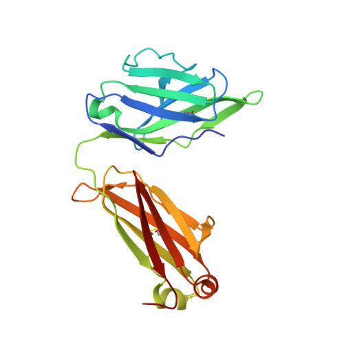
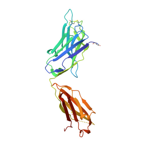
A potent and broad neutralizing antibody recognizes and penetrates the HIV glycan shield.

Pejchal, R.Doores, K.J.Walker, L.M.Khayat, R.Huang, P.S.Wang, S.K.Stanfield, R.L.Julien, J.P.Ramos, A.Crispin, M.Depetris, R.Katpally, U.Marozsan, A.Cupo, A.Maloveste, S.Liu, Y.McBride, R.Ito, Y.Sanders, R.W.Ogohara, C.Paulson, J.C.Feizi, T.Scanlan, C.N.Wong, C.H.Moore, J.P.Olson, W.C.Ward, A.B.Poignard, P.Schief, W.R.Burton, D.R.Wilson, I.A.

(2011) Science 334: 1097-1103

  • DOI: https://doi.org/10.1126/science.1213256
  • Primary Citation of Related Structures:  
    3TV3, 3TWC, 3TYG

  • PubMed Abstract: 

    The HIV envelope (Env) protein gp120 is protected from antibody recognition by a dense glycan shield. However, several of the recently identified PGT broadly neutralizing antibodies appear to interact directly with the HIV glycan coat. Crystal structures of antigen-binding fragments (Fabs) PGT 127 and 128 with Man(9) at 1.65 and 1.29 angstrom resolution, respectively, and glycan binding data delineate a specific high mannose-binding site. Fab PGT 128 complexed with a fully glycosylated gp120 outer domain at 3.25 angstroms reveals that the antibody penetrates the glycan shield and recognizes two conserved glycans as well as a short β-strand segment of the gp120 V3 loop, accounting for its high binding affinity and broad specificity. Furthermore, our data suggest that the high neutralization potency of PGT 127 and 128 immunoglobulin Gs may be mediated by cross-linking Env trimers on the viral surface.


  • Organizational Affiliation

    Department of Molecular Biology, Skaggs Institute for Chemical Biology and International AIDS Vaccine Initiative (IAVI) Neutralizing Antibody Center, nhe Scripps Research Institute, La Jolla, CA 92037, USA.


Macromolecules
Find similar proteins by:  (by identity cutoff)  |  3D Structure
Entity ID: 1
MoleculeChains Sequence LengthOrganismDetailsImage
PGT127 light chain, Ig lambda-2 chain C regionsA [auth L]211Homo sapiensMutation(s): 0 
Gene Names: IGLC2
Entity Groups  
Sequence Clusters30% Identity50% Identity70% Identity90% Identity95% Identity100% Identity
Sequence Annotations
Expand
  • Reference Sequence
Find similar proteins by:  (by identity cutoff)  |  3D Structure
Entity ID: 2
MoleculeChains Sequence LengthOrganismDetailsImage
PGT127 heavy chain, Ig gamma-1 chain C regionB [auth H]239Homo sapiensMutation(s): 0 
Gene Names: IGHG1
UniProt & NIH Common Fund Data Resources
Find proteins for P01857 (Homo sapiens)
Explore P01857 
Go to UniProtKB:  P01857
PHAROS:  P01857
GTEx:  ENSG00000211896 
Entity Groups  
Sequence Clusters30% Identity50% Identity70% Identity90% Identity95% Identity100% Identity
UniProt GroupP01857
Sequence Annotations
Expand
  • Reference Sequence
Oligosaccharides

Help

Entity ID: 3
MoleculeChains Length2D Diagram Glycosylation3D Interactions
alpha-D-mannopyranose-(1-2)-alpha-D-mannopyranose-(1-2)-alpha-D-mannopyranose-(1-3)-[alpha-D-mannopyranose-(1-2)-alpha-D-mannopyranose-(1-6)-[alpha-D-mannopyranose-(1-3)]alpha-D-mannopyranose-(1-6)]alpha-D-mannopyranoseC [auth A]8N/A
Glycosylation Resources
GlyTouCan:  G65997KL
GlyCosmos:  G65997KL
Small Molecules
Ligands 1 Unique
IDChains Name / Formula / InChI Key2D Diagram3D Interactions
AML
Query on AML

Download Ideal Coordinates CCD File 
D [auth H]AMYLAMINE
C5 H13 N
DPBLXKKOBLCELK-UHFFFAOYSA-N
Modified Residues  1 Unique
IDChains TypeFormula2D DiagramParent
PCA
Query on PCA
A [auth L]L-PEPTIDE LINKINGC5 H7 N O3GLN
Experimental Data & Validation

Experimental Data

  • Method: X-RAY DIFFRACTION
  • Resolution: 1.65 Å
  • R-Value Free: 0.215 
  • R-Value Work: 0.192 
  • R-Value Observed: 0.193 
  • Space Group: C 1 2 1
Unit Cell:
Length ( Å )Angle ( ˚ )
a = 103.55α = 90
b = 71.6β = 117.02
c = 78.83γ = 90
Software Package:
Software NamePurpose
Blu-Icedata collection
PHASERphasing
PHENIXrefinement
XDSdata reduction
XSCALEdata scaling

Structure Validation

View Full Validation Report


Entry History 

Deposition Data

Revision History  (Full details and data files)

  • Version 1.0: 2011-10-19
    Type: Initial release
  • Version 1.1: 2011-10-26
    Changes: Database references
  • Version 1.2: 2011-12-07
    Changes: Database references
  • Version 1.3: 2017-08-02
    Changes: Refinement description, Source and taxonomy
  • Version 2.0: 2019-12-25
    Changes: Database references, Derived calculations, Polymer sequence
  • Version 3.0: 2020-07-29
    Type: Remediation
    Reason: Carbohydrate remediation
    Changes: Advisory, Atomic model, Data collection, Derived calculations, Structure summary
  • Version 3.1: 2021-04-28
    Changes: Derived calculations, Source and taxonomy, Structure summary
  • Version 3.2: 2023-09-13
    Changes: Data collection, Database references, Refinement description