3TZO

The role of I87 of CYP158A2 in oxidative coupling reaction


Experimental Data Snapshot

  • Method: X-RAY DIFFRACTION
  • Resolution: 1.76 Å
  • R-Value Free: 0.264 
  • R-Value Work: 0.216 
  • R-Value Observed: 0.216 

wwPDB Validation   3D Report Full Report


Ligand Structure Quality Assessment 


This is version 1.3 of the entry. See complete history


Literature

The role of Ile87 of CYP158A2 in oxidative coupling reaction.

Zhao, B.Bellamine, A.Lei, L.Waterman, M.R.

(2012) Arch Biochem Biophys 518: 127-132

  • DOI: https://doi.org/10.1016/j.abb.2011.12.007
  • Primary Citation of Related Structures:  
    3TZO, 5DE9

  • PubMed Abstract: 

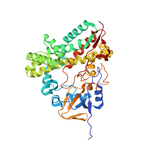
    Both CYP158A1 and CYP158A2 are able to catalyze an oxidative C-C coupling reaction producing biflaviolin or triflaviolin in Streptomyces coelicolor A3(2). The substrate-bound crystal structures of CYP158A2 and CYP158A1 reveal that the side chain of Ile87 in CYP158A2 points to the active site contacting the distal flaviolin molecule, however, the bulkier side chain of Lys90 in CYP158A1 (corresponding to Ile87 in CYP158A2) is toward the distal surface of the protein. These results suggest that these residues could be important in determining product regiospecificity. In order to explore the role of the two residues in catalysis, the reciprocal mutants, Ile87Lys and Lys90Ile, of CYP158A2 and CYP158A1, respectively, were generated and characterized. The mutant Ile87Lys enzyme forms two isomers of biflaviolin instead of three isomers of biflaviolin in wild-type CYP158A2. CYP158A1 containing the substitution of lysine with isoleucine has the same catalytic activity compared with the wild-type CYP158A1. The crystal structure of Ile87Lys showed that the BC loop in the mutant is in a very different orientation compared with the BC loop in both CYP158A1/A2 structures. These results shed light on the mechanism of the oxidative coupling reaction catalyzed by cytochrome P450.


  • Organizational Affiliation

    Department of Biochemistry, Vanderbilt University School of Medicine, Nashville, TN 37232-0146, USA. bin.zhao@vanderbilt.edu


Macromolecules
Find similar proteins by:  (by identity cutoff)  |  3D Structure
Entity ID: 1
MoleculeChains Sequence LengthOrganismDetailsImage
Putative cytochrome P450
A, B
410Streptomyces coelicolorMutation(s): 1 
Gene Names: SCO1207
UniProt
Find proteins for Q9FCA6 (Streptomyces coelicolor (strain ATCC BAA-471 / A3(2) / M145))
Explore Q9FCA6 
Go to UniProtKB:  Q9FCA6
Entity Groups  
Sequence Clusters30% Identity50% Identity70% Identity90% Identity95% Identity100% Identity
UniProt GroupQ9FCA6
Sequence Annotations
Expand
  • Reference Sequence
Experimental Data & Validation

Experimental Data

  • Method: X-RAY DIFFRACTION
  • Resolution: 1.76 Å
  • R-Value Free: 0.264 
  • R-Value Work: 0.216 
  • R-Value Observed: 0.216 
  • Space Group: P 1 21 1
Unit Cell:
Length ( Å )Angle ( ˚ )
a = 59.609α = 90
b = 79.313β = 94.08
c = 87.658γ = 90
Software Package:
Software NamePurpose
HKL-2000data collection
PHASERphasing
CNSrefinement
HKL-2000data reduction
HKL-2000data scaling

Structure Validation

View Full Validation Report


Ligand Structure Quality Assessment 


Entry History 

Deposition Data

Revision History  (Full details and data files)

  • Version 1.0: 2012-01-18
    Type: Initial release
  • Version 1.1: 2012-03-21
    Changes: Database references
  • Version 1.2: 2017-11-08
    Changes: Refinement description
  • Version 1.3: 2024-02-28
    Changes: Data collection, Database references, Derived calculations