


3USD
Crystal Structure of C-lobe of Bovine lactoferrin Complexed with Imidazol (1,2 a) pyridine3-yl-acitic acid at 2.4 A Resolution
- PDB DOI: https://doi.org/10.2210/pdb3USD/pdb
- Classification: METAL BINDING PROTEIN
- Organism(s): Bos taurus
- Mutation(s): No
- Deposited: 2011-11-23 Released: 2011-12-14
Experimental Data Snapshot
- Method: X-RAY DIFFRACTION
- Resolution: 2.40 Å
- R-Value Free: 0.260
- R-Value Work: 0.222
- R-Value Observed: 0.224

This is version 2.1 of the entry. See complete history.

Macromolecules
Find similar proteins by:
(by identity cutoff) | 3D Structure
Entity ID: 1 | |||||
|---|---|---|---|---|---|
| Molecule | Chains | Sequence Length | Organism | Details | Image |
| Lactotransferrin | 335 | Bos taurus | Mutation(s): 0 EC: 3.4.21 | 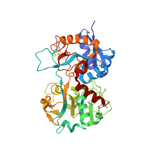 | |
UniProt | |||||
Find proteins for P24627 (Bos taurus) Explore P24627 Go to UniProtKB: P24627 | |||||
Entity Groups | |||||
| Sequence Clusters | 30% Identity50% Identity70% Identity90% Identity95% Identity100% Identity | ||||
| UniProt Group | P24627 | ||||
Sequence AnnotationsExpand | |||||
| |||||
Find similar proteins by: Sequence | 3D Structure
Oligosaccharides


Small Molecules
| Ligands 6 Unique | |||||
|---|---|---|---|---|---|
| ID | Chains | Name / Formula / InChI Key | 2D Diagram | 3D Interactions | |
| NAG Query on NAG | J [auth A] | 2-acetamido-2-deoxy-beta-D-glucopyranose C8 H15 N O6 OVRNDRQMDRJTHS-FMDGEEDCSA-N |  | ||
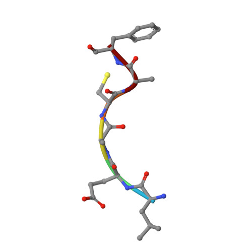
| IDL Query on IDL | K [auth A] | imidazo[1,2-a]pyridin-3-ylacetic acid C9 H8 N2 O2 ZVBVKRNOISRONE-UHFFFAOYSA-N |  | ||
| SO4 Query on SO4 | I [auth A] | SULFATE ION O4 S QAOWNCQODCNURD-UHFFFAOYSA-L |  | ||
| ZN Query on ZN | E [auth A], F [auth A] | ZINC ION Zn PTFCDOFLOPIGGS-UHFFFAOYSA-N |  | ||
| CO3 Query on CO3 | H [auth A] | CARBONATE ION C O3 BVKZGUZCCUSVTD-UHFFFAOYSA-L |  | ||
| FE Query on FE | G [auth A] | FE (III) ION Fe VTLYFUHAOXGGBS-UHFFFAOYSA-N |  | ||
Experimental Data & Validation
Experimental Data
- Method: X-RAY DIFFRACTION
- Resolution: 2.40 Å
- R-Value Free: 0.260
- R-Value Work: 0.222
- R-Value Observed: 0.224
- Space Group: P 1 21 1
Unit Cell:
| Length ( Å ) | Angle ( ˚ ) |
|---|---|
| a = 61.434 | α = 90 |
| b = 50.469 | β = 105.82 |
| c = 64.55 | γ = 90 |
| Software Name | Purpose |
|---|---|
| HKL-2000 | data collection |
| AMoRE | phasing |
| REFMAC | refinement |
| AUTOMAR | data reduction |
| SCALEPACK | data scaling |
Entry History
Deposition Data
- Released Date: 2011-12-14 Deposition Author(s): Shukla, P.K., Gautam, L., Sinha, M., Kaur, P., Sharma, S., Singh, T.P.
Revision History (Full details and data files)
- Version 1.0: 2011-12-14
Type: Initial release - Version 2.0: 2020-07-29
Type: Remediation
Reason: Carbohydrate remediation
Changes: Advisory, Atomic model, Data collection, Database references, Derived calculations, Structure summary - Version 2.1: 2023-11-08
Changes: Data collection, Database references, Refinement description, Structure summary
