3W4U

Human zeta-2 beta-2-s hemoglobin


Experimental Data Snapshot

  • Method: X-RAY DIFFRACTION
  • Resolution: 1.95 Å
  • R-Value Free: 0.255 
  • R-Value Work: 0.205 
  • R-Value Observed: 0.208 

wwPDB Validation   3D Report Full Report


Ligand Structure Quality Assessment 


This is version 1.2 of the entry. See complete history


Literature

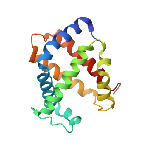
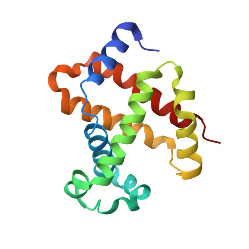
Structure of fully liganded Hb zeta 2 beta 2(s) trapped in a tense conformation

Safo, M.K.Ko, T.-P.Abdulmalik, O.He, Z.Wang, A.H.Schreiter, E.R.Russell, J.E.

(2013) Acta Crystallogr D Biol Crystallogr 69: 2061-2071

  • DOI: https://doi.org/10.1107/S0907444913019197
  • Primary Citation of Related Structures:  
    3W4U

  • PubMed Abstract: 

    A variant Hb ζ2β2(s) that is formed from sickle hemoglobin (Hb S; α2β2(s)) by exchanging adult α-globin with embryonic ζ-globin subunits shows promise as a therapeutic agent for sickle-cell disease (SCD). Hb ζ2β2(s) inhibits the polymerization of deoxygenated Hb S in vitro and reverses characteristic features of SCD in vivo in mouse models of the disorder. When compared with either Hb S or with normal human adult Hb A (α2β2), Hb ζ2β2(s) exhibits atypical properties that include a high oxygen affinity, reduced cooperativity, a weak Bohr effect and blunted 2,3-diphosphoglycerate allostery. Here, the 1.95 Å resolution crystal structure of human Hb ζ2β2(s) that was expressed in complex transgenic knockout mice and purified from their erythrocytes is presented. When fully liganded with carbon monoxide, Hb ζ2β2(s) displays a central water cavity, a ζ1-β(s)2 (or ζ2-β(s)1) interface, intersubunit salt-bridge/hydrogen-bond interactions, C-terminal βHis146 salt-bridge interactions, and a β-cleft, that are highly unusual for a relaxed hemoglobin structure and are more typical of a tense conformation. These quaternary tense-like features contrast with the tertiary relaxed-like conformations of the ζ1β(s)1 dimer and the CD and FG corners, as well as the overall structures of the heme cavities. This crystallographic study provides insights into the altered oxygen-transport properties of Hb ζ2β2(s) and, moreover, decouples tertiary- and quaternary-structural events that are critical to Hb ligand binding and allosteric function.


  • Organizational Affiliation

    Institute for Structural Biology and Drug Discovery, and the Department of Medicinal Chemistry, School of Pharmacy, Virginia Commonwealth University, Richmond, VA 23298, USA.


Macromolecules
Find similar proteins by:  (by identity cutoff)  |  3D Structure
Entity ID: 1
MoleculeChains Sequence LengthOrganismDetailsImage
Hemoglobin subunit zeta
A, C, E
142Homo sapiensMutation(s): 0 
Gene Names: HBZHBZ2
UniProt & NIH Common Fund Data Resources
Find proteins for P02008 (Homo sapiens)
Explore P02008 
Go to UniProtKB:  P02008
PHAROS:  P02008
GTEx:  ENSG00000130656 
Entity Groups  
Sequence Clusters30% Identity50% Identity70% Identity90% Identity95% Identity100% Identity
UniProt GroupP02008
Sequence Annotations
Expand
  • Reference Sequence
Find similar proteins by:  (by identity cutoff)  |  3D Structure
Entity ID: 2
MoleculeChains Sequence LengthOrganismDetailsImage
Hemoglobin subunit beta
B, D, F
146Homo sapiensMutation(s): 1 
Gene Names: HBB
UniProt & NIH Common Fund Data Resources
Find proteins for P68871 (Homo sapiens)
Explore P68871 
Go to UniProtKB:  P68871
PHAROS:  P68871
GTEx:  ENSG00000244734 
Entity Groups  
Sequence Clusters30% Identity50% Identity70% Identity90% Identity95% Identity100% Identity
UniProt GroupP68871
Sequence Annotations
Expand
  • Reference Sequence
Small Molecules
Ligands 2 Unique
IDChains Name / Formula / InChI Key2D Diagram3D Interactions
HEM
Query on HEM

Download Ideal Coordinates CCD File 
G [auth A]
I [auth B]
K [auth C]
M [auth D]
O [auth E]
G [auth A],
I [auth B],
K [auth C],
M [auth D],
O [auth E],
Q [auth F]
PROTOPORPHYRIN IX CONTAINING FE
C34 H32 Fe N4 O4
KABFMIBPWCXCRK-RGGAHWMASA-L
CMO
Query on CMO

Download Ideal Coordinates CCD File 
H [auth A]
J [auth B]
L [auth C]
N [auth D]
P [auth E]
H [auth A],
J [auth B],
L [auth C],
N [auth D],
P [auth E],
R [auth F]
CARBON MONOXIDE
C O
UGFAIRIUMAVXCW-UHFFFAOYSA-N
Experimental Data & Validation

Experimental Data

  • Method: X-RAY DIFFRACTION
  • Resolution: 1.95 Å
  • R-Value Free: 0.255 
  • R-Value Work: 0.205 
  • R-Value Observed: 0.208 
  • Space Group: P 31 2 1
Unit Cell:
Length ( Å )Angle ( ˚ )
a = 115.54α = 90
b = 115.54β = 90
c = 140.9γ = 120
Software Package:
Software NamePurpose
CrystalCleardata collection
CNSrefinement
d*TREKdata reduction
d*TREKdata scaling
CNSphasing

Structure Validation

View Full Validation Report


Ligand Structure Quality Assessment 


Entry History 

Deposition Data

Revision History  (Full details and data files)

  • Version 1.0: 2013-02-06
    Type: Initial release
  • Version 1.1: 2013-10-23
    Changes: Database references
  • Version 1.2: 2023-11-08
    Changes: Data collection, Database references, Derived calculations, Refinement description