4AX7

Hypocrea jecorina Cel6A D221A mutant soaked with 4-Methylumbelliferyl- beta-D-cellobioside


Experimental Data Snapshot

  • Method: X-RAY DIFFRACTION
  • Resolution: 1.70 Å
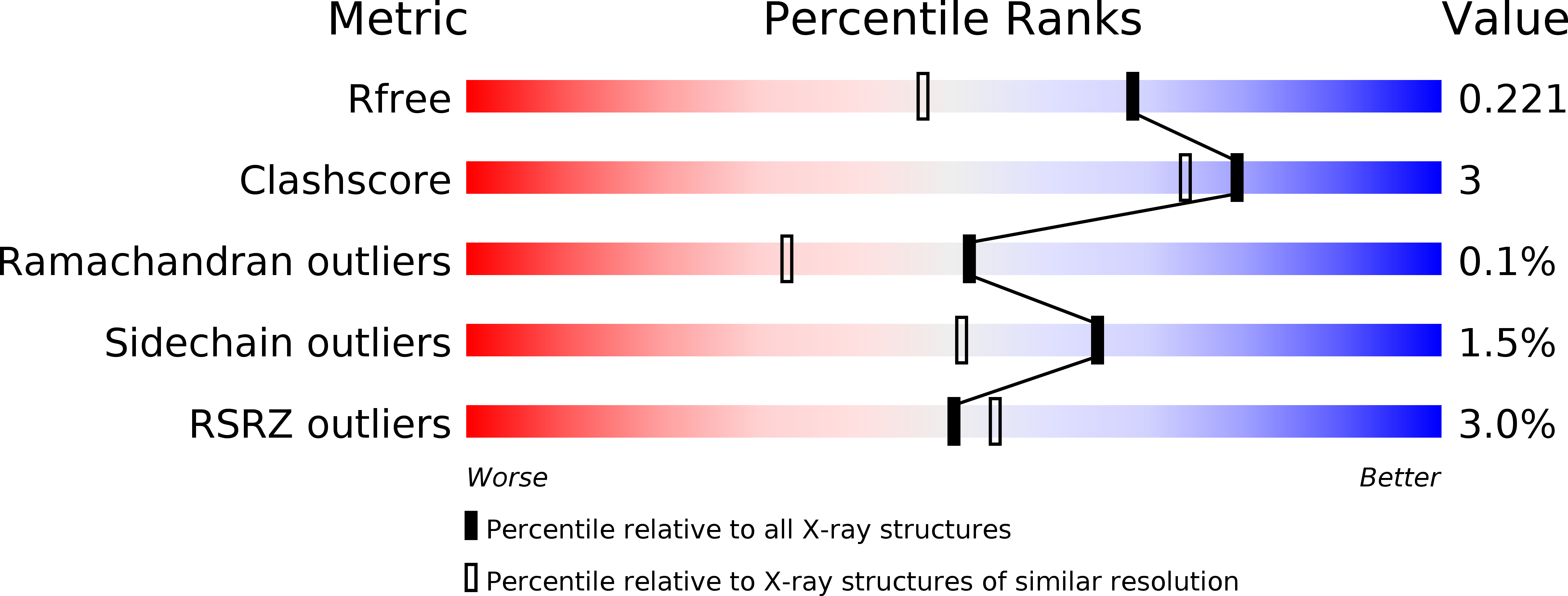
  • R-Value Free: 0.221 
  • R-Value Work: 0.189 
  • R-Value Observed: 0.190 

wwPDB Validation   3D Report Full Report


Ligand Structure Quality Assessment 


This is version 2.1 of the entry. See complete history


Literature

Rational Design, Synthesis, Evaluation and Enzyme-Substrate Structures of Improved Fluorogenic Substrates for Family 6 Glycoside Hydrolases.

Wu, M.Nerinckx, W.Piens, K.Ishida, T.Hansson, H.Sandgren, M.Stahlberg, J.

(2013) FEBS J 280: 184

  • DOI: https://doi.org/10.1111/febs.12060
  • Primary Citation of Related Structures:  
    4AU0, 4AX6, 4AX7

  • PubMed Abstract: 

    Methylumbelliferyl-β-cellobioside (MUF-G2) is a convenient fluorogenic substrate for certain β-glycoside hydrolases (GH). However, hydrolysis of the aglycone is poor with GH family 6 enzymes (GH6), despite strong binding. Prediction of the orientation of the aglycone of MUF-G2 in the +1 subsite of Hypocrea jecorina Cel6A by automated docking suggested umbelliferyl modifications at C4 and C6 for improved recognition. Four modified umbelliferyl-β-cellobiosides [6-chloro-4-methyl- (ClMUF); 6-chloro-4-trifluoromethyl- (ClF3MUF); 4-phenyl- (PhUF); 6-chloro-4-phenyl- (ClPhUF)] were synthesized and tested with GH6, GH7, GH9, GH5 and GH45 cellulases. Indeed the rate of aglycone release by H. jecorina Cel6A was 10-150 times higher than with MUF-G2, although it was still three orders of magnitude lower than with H. jecorina Cel7B. The 4-phenyl substitution drastically reduced the fluorescence intensity of the free aglycone, while ClMUF-G2 could be used for determination of k(cat) and K(M) for H. jecorina Cel6A and Thermobifida fusca Cel6A. Crystal structures of H. jecorina Cel6A D221A mutant soaked with the MUF-, ClMUF- and ClPhUF-β-cellobioside substrates show that the modifications turned the umbelliferyl group 'upside down', with the glycosidic bond better positioned for protonation than with MUF-G2.


  • Organizational Affiliation

    Department of Molecular Biology, Swedish University of Agricultural Sciences, Uppsala, Sweden.


Macromolecules
Find similar proteins by:  (by identity cutoff)  |  3D Structure
Entity ID: 1
MoleculeChains Sequence LengthOrganismDetailsImage
EXOGLUCANASE 2
A, B, C, D
363Trichoderma reeseiMutation(s): 1 
EC: 3.2.1.91
UniProt
Find proteins for P07987 (Hypocrea jecorina)
Explore P07987 
Go to UniProtKB:  P07987
Entity Groups  
Sequence Clusters30% Identity50% Identity70% Identity90% Identity95% Identity100% Identity
UniProt GroupP07987
Sequence Annotations
Expand
  • Reference Sequence
Oligosaccharides

Help

Entity ID: 2
MoleculeChains Length2D Diagram Glycosylation3D Interactions
beta-D-glucopyranose-(1-4)-beta-D-glucopyranose-(1-4)-beta-D-glucopyranose-(1-4)-beta-D-glucopyranose
E, G
4N/A
Glycosylation Resources
GlyTouCan:  G00025MO
GlyCosmos:  G00025MO
GlyGen:  G00025MO
Entity ID: 3
MoleculeChains Length2D Diagram Glycosylation3D Interactions
beta-D-glucopyranose-(1-4)-beta-D-glucopyranose
F, H
2N/A
Glycosylation Resources
GlyTouCan:  G84824ZO
GlyCosmos:  G84824ZO
Small Molecules
Ligands 3 Unique
IDChains Name / Formula / InChI Key2D Diagram3D Interactions
NAG
Query on NAG

Download Ideal Coordinates CCD File 
CA [auth C]
DA [auth C]
I [auth A]
J [auth A]
LA [auth D]
CA [auth C],
DA [auth C],
I [auth A],
J [auth A],
LA [auth D],
MA [auth D],
S [auth B],
T [auth B]
2-acetamido-2-deoxy-beta-D-glucopyranose
C8 H15 N O6
OVRNDRQMDRJTHS-FMDGEEDCSA-N
MAN
Query on MAN

Download Ideal Coordinates CCD File 
AA [auth B]
EA [auth C]
FA [auth C]
GA [auth C]
HA [auth C]
AA [auth B],
EA [auth C],
FA [auth C],
GA [auth C],
HA [auth C],
IA [auth C],
JA [auth C],
K [auth A],
L [auth A],
M [auth A],
N [auth A],
NA [auth D],
O [auth A],
OA [auth D],
P [auth A],
PA [auth D],
Q [auth A],
QA [auth D],
RA [auth D],
SA [auth D],
TA [auth D],
U [auth B],
V [auth B],
W [auth B],
X [auth B],
Y [auth B],
Z [auth B]
alpha-D-mannopyranose
C6 H12 O6
WQZGKKKJIJFFOK-PQMKYFCFSA-N
4MU
Query on 4MU

Download Ideal Coordinates CCD File 
BA [auth B],
KA [auth C],
R [auth A],
UA [auth D]
7-hydroxy-4-methyl-2H-chromen-2-one
C10 H8 O3
HSHNITRMYYLLCV-UHFFFAOYSA-N
Experimental Data & Validation

Experimental Data

  • Method: X-RAY DIFFRACTION
  • Resolution: 1.70 Å
  • R-Value Free: 0.221 
  • R-Value Work: 0.189 
  • R-Value Observed: 0.190 
  • Space Group: P 1
Unit Cell:
Length ( Å )Angle ( ˚ )
a = 48.819α = 103.97
b = 74.679β = 103.43
c = 93.295γ = 90
Software Package:
Software NamePurpose
REFMACrefinement
XDSdata reduction
SCALAdata scaling

Structure Validation

View Full Validation Report


Ligand Structure Quality Assessment 


Entry History 

Deposition Data

Revision History  (Full details and data files)

  • Version 1.0: 2013-01-23
    Type: Initial release
  • Version 1.1: 2020-07-01
    Changes: Advisory, Data collection, Derived calculations, Other
  • Version 2.0: 2020-07-29
    Type: Remediation
    Reason: Carbohydrate remediation
    Changes: Advisory, Atomic model, Data collection, Derived calculations, Structure summary
  • Version 2.1: 2023-12-20
    Changes: Data collection, Database references, Refinement description, Structure summary