4BMY

Structure of futalosine hydrolase mutant of Helicobacter pylori strain 26695


Experimental Data Snapshot

  • Method: X-RAY DIFFRACTION
  • Resolution: 1.65 Å
  • R-Value Free: 0.201 
  • R-Value Work: 0.169 
  • R-Value Observed: 0.171 

wwPDB Validation   3D Report Full Report


This is version 1.3 of the entry. See complete history


Literature

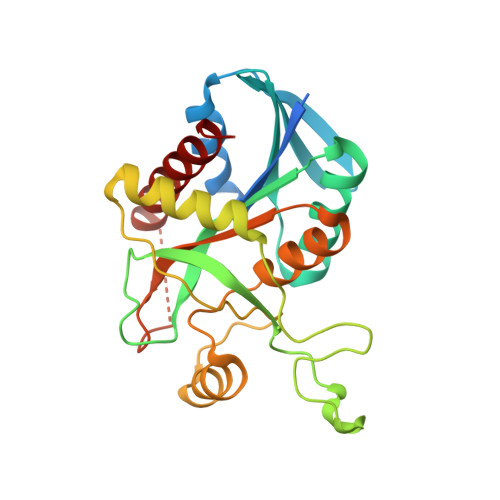
Structural Enzymology of Helicobacter Pylori Methylthioadenosine Nucleosidase in the Futalosine Pathway

Kim, R.Q.Offen, W.A.Davies, G.J.Stubbs, K.A.

(2014) Acta Crystallogr D Biol Crystallogr 70: 177

  • DOI: https://doi.org/10.1107/S1399004713026655
  • Primary Citation of Related Structures:  
    4BMX, 4BMY, 4BMZ, 4BN0

  • PubMed Abstract: 

    The recently discovered futalosine pathway is a promising target for the development of new antibiotics. The enzymes involved in this pathway are crucial for the biosynthesis of the essential prokaryotic respiratory compound menaquinone, and as the pathway is limited to few bacterial species such as the gastric pathogen Helicobacter pylori it is a potential target for specific antibiotics. In this report, the crystal structure of an H. pylori methylthioadenosine nucleosidase (MTAN; an enzyme with broad specificity and activity towards 6-amino-6-deoxyfutalosine), which is involved in the second step of menaquinone biosynthesis, has been elucidated at a resolution of 1.76 Å and refined with R factors of Rwork = 17% and Rfree = 21%. Activity studies on the wild type and active-site mutants show that the hydrolysis of 6-amino-6-deoxyfutalosine follows a mechanism similar to that of Escherichia coli MTAN. Further evidence for this mode of action is supplied by the crystal structures of active-site mutants. Through the use of reaction intermediates, the structures give additional evidence for the previously proposed nucleosidase mechanism. These structures and the confirmed reaction mechanism will provide a structural basis for the design of new inhibitors targeting the futalosine pathway.


  • Organizational Affiliation

    York Structural Biology Laboratory, Department of Chemistry, University of York, York YO10 5DD, England.


Macromolecules
Find similar proteins by:  (by identity cutoff)  |  3D Structure
Entity ID: 1
MoleculeChains Sequence LengthOrganismDetailsImage
MTA/SAH NUCLEOSIDASE
A, B
251Helicobacter pylori 26695Mutation(s): 1 
EC: 3.2.2.9
UniProt
Find proteins for O24915 (Helicobacter pylori (strain ATCC 700392 / 26695))
Explore O24915 
Go to UniProtKB:  O24915
Entity Groups  
Sequence Clusters30% Identity50% Identity70% Identity90% Identity95% Identity100% Identity
UniProt GroupO24915
Sequence Annotations
Expand
  • Reference Sequence
Small Molecules
Ligands 1 Unique
IDChains Name / Formula / InChI Key2D Diagram3D Interactions
SO4
Query on SO4

Download Ideal Coordinates CCD File 
C [auth A]SULFATE ION
O4 S
QAOWNCQODCNURD-UHFFFAOYSA-L
Experimental Data & Validation

Experimental Data

  • Method: X-RAY DIFFRACTION
  • Resolution: 1.65 Å
  • R-Value Free: 0.201 
  • R-Value Work: 0.169 
  • R-Value Observed: 0.171 
  • Space Group: C 2 2 21
Unit Cell:
Length ( Å )Angle ( ˚ )
a = 54.849α = 90
b = 123.414β = 90
c = 145.853γ = 90
Software Package:
Software NamePurpose
REFMACrefinement
XDSdata reduction
SCALAdata scaling
PHASERphasing

Structure Validation

View Full Validation Report


Entry History 

Deposition Data

Revision History  (Full details and data files)

  • Version 1.0: 2013-09-11
    Type: Initial release
  • Version 1.1: 2014-01-15
    Changes: Database references
  • Version 1.2: 2014-01-22
    Changes: Database references
  • Version 1.3: 2023-12-20
    Changes: Data collection, Database references, Derived calculations, Other, Refinement description