4DHL

Crystal structure of red kidney bean purple acid phosphatase in complex with Maybridge fragment MO07123


Experimental Data Snapshot

  • Method: X-RAY DIFFRACTION
  • Resolution: 2.30 Å
  • R-Value Free: 0.206 
  • R-Value Work: 0.158 
  • R-Value Observed: 0.160 

wwPDB Validation   3D Report Full Report


Ligand Structure Quality Assessment 


This is version 2.0 of the entry. See complete history


Literature

Identification of purple acid phosphatase inhibitors by fragment-based screening: promising new leads for osteoporosis therapeutics.

Feder, D.Hussein, W.M.Clayton, D.J.Kan, M.W.Schenk, G.McGeary, R.P.Guddat, L.W.

(2012) Chem Biol Drug Des 80: 665-674

  • DOI: https://doi.org/10.1111/cbdd.12001
  • Primary Citation of Related Structures:  
    4DHL, 4DSY, 4DT2

  • PubMed Abstract: 

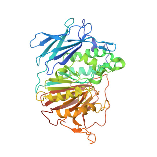
    Purple acid phosphatases are metalloenzymes found in animals, plants and fungi. They possess a binuclear metal centre to catalyse the hydrolysis of phosphate esters and anhydrides under acidic conditions. In humans, elevated purple acid phosphatases levels in sera are correlated with the progression of osteoporosis and metabolic bone malignancies, making this enzyme a target for the development of new chemotherapeutics to treat bone-related illnesses. To date, little progress has been achieved towards the design of specific and potent inhibitors of this enzyme that have drug-like properties. Here, we have undertaken a fragment-based screening approach using a 500-compound library identifying three inhibitors of purple acid phosphatases with K(i) values in the 30-60 μm range. Ligand efficiency values are 0.39-0.44 kcal/mol per heavy atom. X-ray crystal structures of these compounds in complex with a plant purple acid phosphatases (2.3-2.7 Å resolution) have been determined and show that all bind in the active site within contact of the binuclear centre. For one of these compounds, the phenyl ring is positioned within 3.5 Å of the binuclear centre. Docking simulations indicate that the three compounds fit into the active site of human purple acid phosphatases. These studies open the way to the design of more potent and selective inhibitors of purple acid phosphatases that can be tested as anti-osteoporotic drug leads.


  • Organizational Affiliation

    School of Chemistry and Molecular Biosciences, The University of Queensland, Brisbane, Qld 4072, Australia.


Macromolecules
Find similar proteins by:  (by identity cutoff)  |  3D Structure
Entity ID: 1
MoleculeChains Sequence LengthOrganismDetailsImage
Purple acid phosphataseA,
B,
C [auth D],
D [auth C]
426Phaseolus vulgarisMutation(s): 0 
EC: 3.1.3.2
UniProt
Find proteins for P80366 (Phaseolus vulgaris)
Explore P80366 
Go to UniProtKB:  P80366
Entity Groups  
Sequence Clusters30% Identity50% Identity70% Identity90% Identity95% Identity100% Identity
UniProt GroupP80366
Sequence Annotations
Expand
  • Reference Sequence
Oligosaccharides

Help

Entity ID: 2
MoleculeChains Length2D Diagram Glycosylation3D Interactions
alpha-L-fucopyranose-(1-3)-[2-acetamido-2-deoxy-beta-D-glucopyranose-(1-4)]2-acetamido-2-deoxy-beta-D-glucopyranose
E, F, G, I, J
E, F, G, I, J, M
3N-Glycosylation
Glycosylation Resources
GlyTouCan:  G11971MR
GlyCosmos:  G11971MR
GlyGen:  G11971MR
Entity ID: 3
MoleculeChains Length2D Diagram Glycosylation3D Interactions
alpha-L-fucopyranose-(1-3)-2-acetamido-2-deoxy-beta-D-glucopyranose
H, K, L
2N-Glycosylation
Glycosylation Resources
GlyTouCan:  G80587NA
GlyCosmos:  G80587NA
GlyGen:  G80587NA
Small Molecules
Ligands 7 Unique
IDChains Name / Formula / InChI Key2D Diagram3D Interactions
NAG
Query on NAG

Download Ideal Coordinates CCD File 
AA [auth B]
JA [auth D]
MA [auth D]
SA [auth C]
TA [auth C]
AA [auth B],
JA [auth D],
MA [auth D],
SA [auth C],
TA [auth C],
U [auth A],
Z [auth B]
2-acetamido-2-deoxy-beta-D-glucopyranose
C8 H15 N O6
OVRNDRQMDRJTHS-FMDGEEDCSA-N
0K7
Query on 0K7

Download Ideal Coordinates CCD File 
DA [auth D],
P [auth A],
PA [auth C],
X [auth B]
2-(4-methylphenyl)-1,3-thiazole-4-carboxylic acid
C11 H9 N O2 S
MTVWIZYQYPRZGZ-UHFFFAOYSA-N
SO4
Query on SO4

Download Ideal Coordinates CCD File 
FA [auth D],
R [auth A],
RA [auth C],
S [auth A],
Y [auth B]
SULFATE ION
O4 S
QAOWNCQODCNURD-UHFFFAOYSA-L
GOL
Query on GOL

Download Ideal Coordinates CCD File 
EA [auth D],
Q [auth A],
QA [auth C]
GLYCEROL
C3 H8 O3
PEDCQBHIVMGVHV-UHFFFAOYSA-N
ZN
Query on ZN

Download Ideal Coordinates CCD File 
BA [auth D],
N [auth A],
NA [auth C],
V [auth B]
ZINC ION
Zn
PTFCDOFLOPIGGS-UHFFFAOYSA-N
EDO
Query on EDO

Download Ideal Coordinates CCD File 
GA [auth D]
HA [auth D]
IA [auth D]
KA [auth D]
LA [auth D]
GA [auth D],
HA [auth D],
IA [auth D],
KA [auth D],
LA [auth D],
T [auth A]
1,2-ETHANEDIOL
C2 H6 O2
LYCAIKOWRPUZTN-UHFFFAOYSA-N
FE
Query on FE

Download Ideal Coordinates CCD File 
CA [auth D],
O [auth A],
OA [auth C],
W [auth B]
FE (III) ION
Fe
VTLYFUHAOXGGBS-UHFFFAOYSA-N
Binding Affinity Annotations 
IDSourceBinding Affinity
0K7 Binding MOAD:  4DHL Ki: 3.30e+4 (nM) from 1 assay(s)
PDBBind:  4DHL Ki: 3.30e+4 (nM) from 1 assay(s)
Experimental Data & Validation

Experimental Data

  • Method: X-RAY DIFFRACTION
  • Resolution: 2.30 Å
  • R-Value Free: 0.206 
  • R-Value Work: 0.158 
  • R-Value Observed: 0.160 
  • Space Group: P 31 2 1
Unit Cell:
Length ( Å )Angle ( ˚ )
a = 126.097α = 90
b = 126.097β = 90
c = 297.98γ = 120
Software Package:
Software NamePurpose
PHENIXrefinement

Structure Validation

View Full Validation Report


Ligand Structure Quality Assessment 


Entry History 

Deposition Data

Revision History  (Full details and data files)

  • Version 1.0: 2012-12-12
    Type: Initial release
  • Version 1.1: 2013-01-02
    Changes: Database references
  • Version 2.0: 2020-07-29
    Type: Remediation
    Reason: Carbohydrate remediation
    Changes: Advisory, Atomic model, Data collection, Derived calculations, Structure summary