4DO1

The crystal structures of 4-methoxybenzoate bound CYP199A4


Experimental Data Snapshot

  • Method: X-RAY DIFFRACTION
  • Resolution: 2.00 Å
  • R-Value Free: 0.189 
  • R-Value Work: 0.154 
  • R-Value Observed: 0.155 

wwPDB Validation   3D Report Full Report


Ligand Structure Quality Assessment 


This is version 1.1 of the entry. See complete history


Literature

The crystal structures of 4-methoxybenzoate bound CYP199A2 and CYP199A4: structural changes on substrate binding and the identification of an anion binding site

Bell, S.G.Yang, W.Tan, A.B.H.Zhou, R.Johnson, E.O.D.Zhang, A.Zhou, W.Rao, Z.Wong, L.-L.

(2012) Dalton Trans 41: 8703-8714

  • DOI: https://doi.org/10.1039/c2dt30783a
  • Primary Citation of Related Structures:  
    4DNJ, 4DNZ, 4DO1

  • PubMed Abstract: 

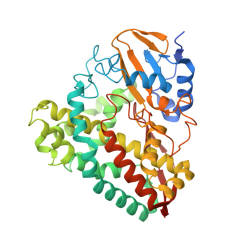
    The crystal structures of the 4-methoxybenzoate bound forms of cytochrome P450 enzymes CYP199A2 and CYP199A4 from the Rhodopseudomonas palustris strains CGA009 and HaA2 have been solved. The structures of these two enzymes, which share 86% sequence identity, are very similar though some differences are found on the proximal surface. In these structures the enzymes have a closed conformation, in contrast to the substrate-free form of CYP199A2 where an obvious substrate access channel is observed. The switch from an open to a closed conformation arises from pronounced residue side-chain movements and alterations of ion pair and hydrogen bonding interactions at the entrance of the access channel. A chloride ion bound just inside the protein surface caps the entrance to the active site and protects the substrate and the heme from the external solvent. In both structures the substrate is held in place via hydrophobic and hydrogen bond interactions. The methoxy group is located over the heme iron, accounting for the high activity and selectivity of these enzymes for oxidative demethylation of the substrate. Mutagenesis studies on CYP199A4 highlight the involvement of hydrophobic (Phe185) and hydrophilic (Arg92, Ser95 and Arg243) amino acid residues in the binding of para-substituted benzoates by these enzymes.


  • Organizational Affiliation

    Department of Chemistry, University of Oxford, Inorganic Chemistry Laboratory, South Parks Road, Oxford, OX1 3QR, UK. stephen.bell@adelaide.edu.au


Macromolecules
Find similar proteins by:  (by identity cutoff)  |  3D Structure
Entity ID: 1
MoleculeChains Sequence LengthOrganismDetailsImage
Cytochrome P450
A, B, C, D
410Rhodopseudomonas palustris HaA2Mutation(s): 0 
Gene Names: RPB_3613
UniProt
Find proteins for Q2IU02 (Rhodopseudomonas palustris (strain HaA2))
Explore Q2IU02 
Go to UniProtKB:  Q2IU02
Entity Groups  
Sequence Clusters30% Identity50% Identity70% Identity90% Identity95% Identity100% Identity
UniProt GroupQ2IU02
Sequence Annotations
Expand
  • Reference Sequence
Small Molecules
Ligands 5 Unique
IDChains Name / Formula / InChI Key2D Diagram3D Interactions
HEM
Query on HEM

Download Ideal Coordinates CCD File 
E [auth A],
J [auth B],
P [auth C],
U [auth D]
PROTOPORPHYRIN IX CONTAINING FE
C34 H32 Fe N4 O4
KABFMIBPWCXCRK-RGGAHWMASA-L
ANN
Query on ANN

Download Ideal Coordinates CCD File 
F [auth A],
K [auth B],
Q [auth C],
V [auth D]
4-METHOXYBENZOIC ACID
C8 H8 O3
ZEYHEAKUIGZSGI-UHFFFAOYSA-N
SO4
Query on SO4

Download Ideal Coordinates CCD File 
G [auth A],
L [auth B],
M [auth B],
R [auth C],
W [auth D]
SULFATE ION
O4 S
QAOWNCQODCNURD-UHFFFAOYSA-L
GOL
Query on GOL

Download Ideal Coordinates CCD File 
H [auth A],
N [auth B],
S [auth C],
X [auth D]
GLYCEROL
C3 H8 O3
PEDCQBHIVMGVHV-UHFFFAOYSA-N
CL
Query on CL

Download Ideal Coordinates CCD File 
I [auth A],
O [auth B],
T [auth C],
Y [auth D]
CHLORIDE ION
Cl
VEXZGXHMUGYJMC-UHFFFAOYSA-M
Binding Affinity Annotations 
IDSourceBinding Affinity
ANN Binding MOAD:  4DO1 Kd: 280 (nM) from 1 assay(s)
Experimental Data & Validation

Experimental Data

  • Method: X-RAY DIFFRACTION
  • Resolution: 2.00 Å
  • R-Value Free: 0.189 
  • R-Value Work: 0.154 
  • R-Value Observed: 0.155 
  • Space Group: P 21 21 21
Unit Cell:
Length ( Å )Angle ( ˚ )
a = 107.021α = 90
b = 143.479β = 90
c = 172.768γ = 90
Software Package:
Software NamePurpose
HKL-2000data collection
CCP4model building
REFMACrefinement
HKL-2000data reduction
HKL-2000data scaling
CCP4phasing

Structure Validation

View Full Validation Report


Ligand Structure Quality Assessment 


Entry History 

Deposition Data

Revision History  (Full details and data files)

  • Version 1.0: 2012-08-15
    Type: Initial release
  • Version 1.1: 2023-11-08
    Changes: Data collection, Database references, Derived calculations, Refinement description