


High resolution structure of Notexin
Ullah, A., Spencer, P., Murakami, M.T., Arni, R.K.To be published.
Experimental Data Snapshot
wwPDB Validation 3D Report Full Report

Entity ID: 1 | |||||
|---|---|---|---|---|---|
| Molecule | Chains | Sequence Length | Organism | Details | Image |
| Phospholipase A2 | 119 | Notechis scutatus scutatus | Mutation(s): 0 EC: 3.1.1.4 | 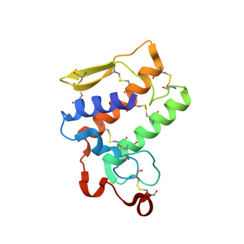 | |
UniProt | |||||
Find proteins for P00608 (Notechis scutatus scutatus) Explore P00608 Go to UniProtKB: P00608 | |||||
Entity Groups | |||||
| Sequence Clusters | 30% Identity50% Identity70% Identity90% Identity95% Identity100% Identity | ||||
| UniProt Group | P00608 | ||||
Sequence AnnotationsExpand | |||||
| |||||
| Ligands 2 Unique | |||||
|---|---|---|---|---|---|
| ID | Chains | Name / Formula / InChI Key | 2D Diagram | 3D Interactions | |
| MES Query on MES | D [auth A] | 2-(N-MORPHOLINO)-ETHANESULFONIC ACID C6 H13 N O4 S SXGZJKUKBWWHRA-UHFFFAOYSA-N |  | ||
| SO4 Query on SO4 | B [auth A], C [auth A] | SULFATE ION O4 S QAOWNCQODCNURD-UHFFFAOYSA-L |  | ||
| Length ( Å ) | Angle ( ˚ ) |
|---|---|
| a = 74.1 | α = 90 |
| b = 74.1 | β = 90 |
| c = 48.738 | γ = 120 |
| Software Name | Purpose |
|---|---|
| NatXray | data collection |
| MOLREP | phasing |
| REFMAC | refinement |
| DENZO | data reduction |
| SCALEPACK | data scaling |
RCSB PDB (citation) is hosted by
RCSB PDB is a member of the

