4EGM

The X-ray crystal structure of CYP199A4 in complex with 4-ethylbenzoic acid


Experimental Data Snapshot

  • Method: X-RAY DIFFRACTION
  • Resolution: 2.91 Å
  • R-Value Free: 0.249 
  • R-Value Work: 0.182 
  • R-Value Observed: 0.185 

wwPDB Validation   3D Report Full Report


Ligand Structure Quality Assessment 


This is version 1.1 of the entry. See complete history


Literature

Investigation of the substrate range of CYP199A4: modification of the partition between hydroxylation and desaturation activities by substrate and protein engineering

Bell, S.G.Zhou, R.M.Yang, W.Tan, A.B.H.Gentleman, A.S.Wong, L.-L.Zhou, W.

(2012) Chemistry 18: 16677-16688

  • DOI: https://doi.org/10.1002/chem.201202776
  • Primary Citation of Related Structures:  
    4EGM, 4EGN, 4EGO, 4EGP

  • PubMed Abstract: 

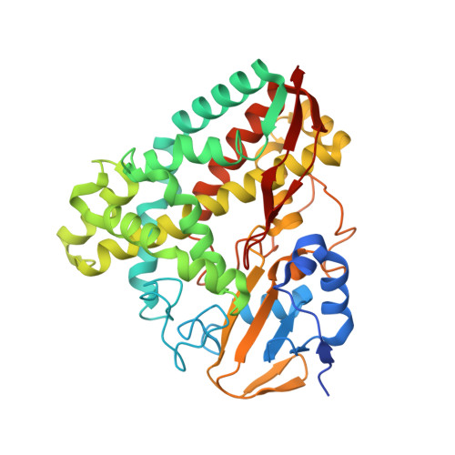
    The cytochrome P450 enzyme CYP199A4, from Rhodopseudomonas palustris HaA2, can efficiently demethylate 4-methoxybenzoic acid. It is also capable of oxidising a range of other related substrates. By investigating substrates with different substituents and ring systems we have been able to show that the carboxylate group and the nature of the ring system and the substituent are all important for optimal substrate binding and activity. The structures of the veratric acid, 2-naphthoic acid and indole-6-carboxylic acid substrate-bound CYP199A4 complexes reveal the substrate binding modes and the side-chain conformational changes of the active site residues to accommodate these larger substrates. They also provide a rationale for the selectivity of product oxidation. The oxidation of alkyl substituted benzoic acids by CYP199A4 is more complex, with desaturation reactions competing with hydroxylation activity. The structure of 4-ethylbenzoic acid-bound CYP199A4 revealed that the substrate is held in a similar position to 4-methoxybenzoic acid, and that the C(β) C-H bonds of the ethyl group are closer to the heme iron than those of the C(α) (3.5 vs. 4.8 Å). This observation, when coupled to the relative energies of the reaction intermediates, indicates that the positioning of the alkyl group relative to the heme iron may be critical in determining the amount of desaturation that is observed. By mutating a single residue in the active site of CYP199A4 (Phe185) we were able to convert the enzyme into a 4-ethylbenzoic acid desaturase.


  • Organizational Affiliation

    Department of Chemistry, University of Oxford, UK. stephen.bell@adelaide.edu.au


Macromolecules
Find similar proteins by:  (by identity cutoff)  |  3D Structure
Entity ID: 1
MoleculeChains Sequence LengthOrganismDetailsImage
Cytochrome P450
A, B, C, D
410Rhodopseudomonas palustris HaA2Mutation(s): 0 
Gene Names: RPB_3613
UniProt
Find proteins for Q2IU02 (Rhodopseudomonas palustris (strain HaA2))
Explore Q2IU02 
Go to UniProtKB:  Q2IU02
Entity Groups  
Sequence Clusters30% Identity50% Identity70% Identity90% Identity95% Identity100% Identity
UniProt GroupQ2IU02
Sequence Annotations
Expand
  • Reference Sequence
Small Molecules
Ligands 5 Unique
IDChains Name / Formula / InChI Key2D Diagram3D Interactions
HEM
Query on HEM

Download Ideal Coordinates CCD File 
BA [auth D],
E [auth A],
M [auth B],
T [auth C]
PROTOPORPHYRIN IX CONTAINING FE
C34 H32 Fe N4 O4
KABFMIBPWCXCRK-RGGAHWMASA-L
EGM
Query on EGM

Download Ideal Coordinates CCD File 
CA [auth D],
F [auth A],
N [auth B],
U [auth C]
4-ethylbenzoic acid
C9 H10 O2
ZQVKTHRQIXSMGY-UHFFFAOYSA-N
SO4
Query on SO4

Download Ideal Coordinates CCD File 
AA [auth C]
FA [auth D]
GA [auth D]
HA [auth D]
I [auth A]
AA [auth C],
FA [auth D],
GA [auth D],
HA [auth D],
I [auth A],
IA [auth D],
J [auth A],
K [auth A],
L [auth A],
Q [auth B],
R [auth B],
S [auth B],
X [auth C],
Y [auth C],
Z [auth C]
SULFATE ION
O4 S
QAOWNCQODCNURD-UHFFFAOYSA-L
GOL
Query on GOL

Download Ideal Coordinates CCD File 
EA [auth D],
H [auth A],
P [auth B],
W [auth C]
GLYCEROL
C3 H8 O3
PEDCQBHIVMGVHV-UHFFFAOYSA-N
CL
Query on CL

Download Ideal Coordinates CCD File 
DA [auth D],
G [auth A],
O [auth B],
V [auth C]
CHLORIDE ION
Cl
VEXZGXHMUGYJMC-UHFFFAOYSA-M
Experimental Data & Validation

Experimental Data

  • Method: X-RAY DIFFRACTION
  • Resolution: 2.91 Å
  • R-Value Free: 0.249 
  • R-Value Work: 0.182 
  • R-Value Observed: 0.185 
  • Space Group: P 21 21 21
Unit Cell:
Length ( Å )Angle ( ˚ )
a = 106.864α = 90
b = 143.279β = 90
c = 172.513γ = 90
Software Package:
Software NamePurpose
HKL-2000data collection
PHASERphasing
REFMACrefinement
HKL-2000data reduction
HKL-2000data scaling

Structure Validation

View Full Validation Report


Ligand Structure Quality Assessment 


Entry History 

Deposition Data

Revision History  (Full details and data files)

  • Version 1.0: 2013-02-20
    Type: Initial release
  • Version 1.1: 2023-11-08
    Changes: Data collection, Database references, Derived calculations, Refinement description