4ENT

Structure of the S234A variant of E. coli KatE


Experimental Data Snapshot

  • Method: X-RAY DIFFRACTION
  • Resolution: 1.70 Å
  • R-Value Free: 0.197 
  • R-Value Work: 0.158 
  • R-Value Observed: 0.160 

wwPDB Validation   3D Report Full Report


Ligand Structure Quality Assessment 


This is version 1.1 of the entry. See complete history


Literature

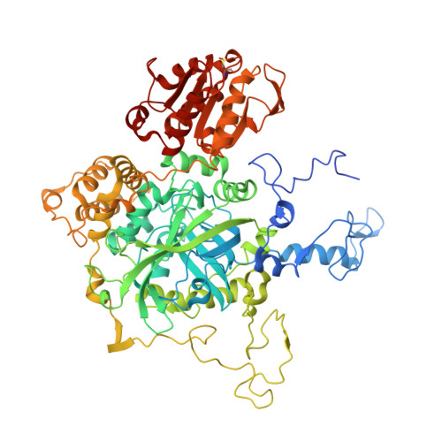
Influence of main channel structure on H(2)O(2) access to the heme cavity of catalase KatE of Escherichia coli.

Jha, V.Chelikani, P.Carpena, X.Fita, I.Loewen, P.C.

(2012) Arch Biochem Biophys 526: 54-59

  • DOI: https://doi.org/10.1016/j.abb.2012.06.010
  • Primary Citation of Related Structures:  
    4ENP, 4ENQ, 4ENR, 4ENS, 4ENT, 4ENU, 4ENV, 4ENW

  • PubMed Abstract: 

    The main channel for H(2)O(2) access to the heme cavity in large subunit catalases is twice as long as in small subunit catalases and is divided into two distinct parts. Like small subunit catalases, the 15Å of the channel adjacent to the heme has a predominantly hydrophobic surface with only weak water occupancy, but the next 15Å extending to the protein surface is hydrophilic and contains a complex water matrix in multiple passages. At the approximate junction of these two sections are a conserved serine and glutamate that are hydrogen bonded and associated with H(2)O(2) in inactive variants. Mutation of these residues changed the dimensions of the channel, both enlarging and constricting it, and also changed the solvent occupancy in the hydrophobic, inner section of the main channel. Despite these structural changes and the prominent location of the residues in the channel, the variants exhibited less than a 2-fold change in the k(cat) and apparent K(M) kinetic constants. These results reflect the importance of the complex multi-passage structure of the main channel. Surprisingly, mutation of either the serine or glutamate to an aliphatic side chain interfered with heme oxidation to heme d.


  • Organizational Affiliation

    Department of Microbiology, University of Manitoba, Winnipeg, MB, Canada.


Macromolecules
Find similar proteins by:  (by identity cutoff)  |  3D Structure
Entity ID: 1
MoleculeChains Sequence LengthOrganismDetailsImage
Catalase HPII
A, B, C, D
753Escherichia coli K-12Mutation(s): 1 
Gene Names: b1732JW1721katE
EC: 1.11.1.6
UniProt
Find proteins for P21179 (Escherichia coli (strain K12))
Explore P21179 
Go to UniProtKB:  P21179
Entity Groups  
Sequence Clusters30% Identity50% Identity70% Identity90% Identity95% Identity100% Identity
UniProt GroupP21179
Sequence Annotations
Expand
  • Reference Sequence
Small Molecules
Modified Residues  1 Unique
IDChains TypeFormula2D DiagramParent
OCS
Query on OCS
A, B, C, D
L-PEPTIDE LINKINGC3 H7 N O5 SCYS
Experimental Data & Validation

Experimental Data

  • Method: X-RAY DIFFRACTION
  • Resolution: 1.70 Å
  • R-Value Free: 0.197 
  • R-Value Work: 0.158 
  • R-Value Observed: 0.160 
  • Space Group: P 1 21 1
Unit Cell:
Length ( Å )Angle ( ˚ )
a = 93.46α = 90
b = 133.05β = 109.58
c = 122.19γ = 90
Software Package:
Software NamePurpose
SCALAdata scaling
REFMACrefinement
PDB_EXTRACTdata extraction

Structure Validation

View Full Validation Report


Ligand Structure Quality Assessment 


Entry History 

Deposition Data

Revision History  (Full details and data files)

  • Version 1.0: 2012-05-02
    Type: Initial release
  • Version 1.1: 2013-02-27
    Changes: Database references