4GIM

Crystal Structure of Pseudouridine Monophosphate Glycosidase Complexed with Pseudouridine 5'-phosphate


Experimental Data Snapshot

  • Method: X-RAY DIFFRACTION
  • Resolution: 1.80 Å
  • R-Value Free: 0.207 
  • R-Value Work: 0.177 
  • R-Value Observed: 0.179 

wwPDB Validation   3D Report Full Report


Ligand Structure Quality Assessment 


This is version 1.2 of the entry. See complete history


Literature

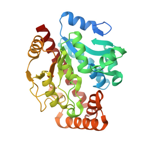
Pseudouridine monophosphate glycosidase: a new glycosidase mechanism.

Huang, S.Mahanta, N.Begley, T.P.Ealick, S.E.

(2012) Biochemistry 51: 9245-9255

  • DOI: https://doi.org/10.1021/bi3006829
  • Primary Citation of Related Structures:  
    4GIJ, 4GIK, 4GIL, 4GIM

  • PubMed Abstract: 

    Pseudouridine (Ψ), the most abundant modification in RNA, is synthesized in situ using Ψ synthase. Recently, a pathway for the degradation of Ψ was described [Preumont, A., Snoussi, K., Stroobant, V., Collet, J. F., and Van Schaftingen, E. (2008) J. Biol. Chem. 283, 25238-25246]. In this pathway, Ψ is first converted to Ψ 5'-monophosphate (ΨMP) by Ψ kinase and then ΨMP is degraded by ΨMP glycosidase to uracil and ribose 5-phosphate. ΨMP glycosidase is the first example of a mechanistically characterized enzyme that cleaves a C-C glycosidic bond. Here we report X-ray crystal structures of Escherichia coli ΨMP glycosidase and a complex of the K166A mutant with ΨMP. We also report the structures of a ring-opened ribose 5-phosphate adduct and a ring-opened ribose ΨMP adduct. These structures provide four snapshots along the reaction coordinate. The structural studies suggested that the reaction utilizes a Lys166 adduct during catalysis. Biochemical and mass spectrometry data further confirmed the existence of a lysine adduct. We used site-directed mutagenesis combined with kinetic analysis to identify roles for specific active site residues. Together, these data suggest that ΨMP glycosidase catalyzes the cleavage of the C-C glycosidic bond through a novel ribose ring-opening mechanism.


  • Organizational Affiliation

    Department of Chemistry and Chemical Biology, Cornell University, Ithaca, New York 14853, United States.


Macromolecules
Find similar proteins by:  (by identity cutoff)  |  3D Structure
Entity ID: 1
MoleculeChains Sequence LengthOrganismDetailsImage
Pseudouridine-5'-phosphate glycosidase
A, B, C
335Escherichia coliMutation(s): 1 
Gene Names: b2165JW2152psuGyeiN
EC: 3.2
UniProt
Find proteins for P33025 (Escherichia coli (strain K12))
Explore P33025 
Go to UniProtKB:  P33025
Entity Groups  
Sequence Clusters30% Identity50% Identity70% Identity90% Identity95% Identity100% Identity
UniProt GroupP33025
Sequence Annotations
Expand
  • Reference Sequence
Experimental Data & Validation

Experimental Data

  • Method: X-RAY DIFFRACTION
  • Resolution: 1.80 Å
  • R-Value Free: 0.207 
  • R-Value Work: 0.177 
  • R-Value Observed: 0.179 
  • Space Group: P 21 21 21
Unit Cell:
Length ( Å )Angle ( ˚ )
a = 60.657α = 90
b = 76.511β = 90
c = 199.162γ = 90
Software Package:
Software NamePurpose
SCALEPACKdata scaling
PHENIXrefinement
PDB_EXTRACTdata extraction
HKL-2000data collection
HKL-2000data reduction
MOLREPphasing

Structure Validation

View Full Validation Report


Ligand Structure Quality Assessment 


Entry History 

Deposition Data

Revision History  (Full details and data files)

  • Version 1.0: 2012-10-31
    Type: Initial release
  • Version 1.1: 2013-01-02
    Changes: Database references
  • Version 1.2: 2023-09-13
    Changes: Data collection, Database references, Derived calculations, Refinement description