


4GIU
Bianthranilate-like analogue bound in inner site of anthranilate phosphoribosyltransferase (AnPRT; trpD).
- PDB DOI: https://doi.org/10.2210/pdb4GIU/pdb
- Classification: TRANSFERASE/TRANSFERASE INHIBITOR
- Organism(s): Mycobacterium tuberculosis
- Expression System: Escherichia coli BL21(DE3)
- Mutation(s): No
- Deposited: 2012-08-09 Released: 2013-08-14
Experimental Data Snapshot
- Method: X-RAY DIFFRACTION
- Resolution: 1.67 Å
- R-Value Free: 0.223
- R-Value Work: 0.199
- R-Value Observed: 0.200

This is version 1.4 of the entry. See complete history.

Macromolecules
Find similar proteins by:
(by identity cutoff) | 3D Structure
Entity ID: 1 | |||||
|---|---|---|---|---|---|
| Molecule | Chains | Sequence Length | Organism | Details | Image |
| Anthranilate phosphoribosyltransferase | 378 | Mycobacterium tuberculosis | Mutation(s): 0 Gene Names: MT2248, MTCY190.03c, Rv2192c, trpD EC: 2.4.2.18 | 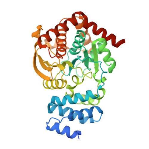 | |
UniProt | |||||
Find proteins for P9WFX5 (Mycobacterium tuberculosis (strain ATCC 25618 / H37Rv)) Explore P9WFX5 Go to UniProtKB: P9WFX5 | |||||
Entity Groups | |||||
| Sequence Clusters | 30% Identity50% Identity70% Identity90% Identity95% Identity100% Identity | ||||
| UniProt Group | P9WFX5 | ||||
Sequence AnnotationsExpand | |||||
| |||||
Small Molecules
| Ligands 5 Unique | |||||
|---|---|---|---|---|---|
| ID | Chains | Name / Formula / InChI Key | 2D Diagram | 3D Interactions | |
| PRP Query on PRP | C [auth A], L [auth B] | 1-O-pyrophosphono-5-O-phosphono-alpha-D-ribofuranose C5 H13 O14 P3 PQGCEDQWHSBAJP-TXICZTDVSA-N |  | ||
| 636 Query on 636 | F [auth A], O [auth B] | 2-[(2-carboxy-5-methylphenyl)amino]-3-methylbenzoic acid C16 H15 N O4 AQDGEUMMMQRBPR-UHFFFAOYSA-N |  | ||
| GOL Query on GOL | G [auth A] H [auth A] I [auth A] P [auth B] Q [auth B] | GLYCEROL C3 H8 O3 PEDCQBHIVMGVHV-UHFFFAOYSA-N |  | ||
| DMS Query on DMS | J [auth A], K [auth A], S [auth B] | DIMETHYL SULFOXIDE C2 H6 O S IAZDPXIOMUYVGZ-UHFFFAOYSA-N |  | ||
| MG Query on MG | D [auth A], E [auth A], M [auth B], N [auth B] | MAGNESIUM ION Mg JLVVSXFLKOJNIY-UHFFFAOYSA-N |  | ||
Experimental Data & Validation
Experimental Data
- Method: X-RAY DIFFRACTION
- Resolution: 1.67 Å
- R-Value Free: 0.223
- R-Value Work: 0.199
- R-Value Observed: 0.200
- Space Group: P 21 21 2
Unit Cell:
| Length ( Å ) | Angle ( ˚ ) |
|---|---|
| a = 111.442 | α = 90 |
| b = 80.752 | β = 90 |
| c = 78.723 | γ = 90 |
| Software Name | Purpose |
|---|---|
| Blu-Ice | data collection |
| PHASER | phasing |
| PHENIX | refinement |
| XDS | data reduction |
| SCALA | data scaling |
Entry History
Deposition Data
- Released Date: 2013-08-14 Deposition Author(s): Evans, G.L., Baker, E.N., Lott, J.S., TB Structural Genomics Consortium (TBSGC)
Revision History (Full details and data files)
- Version 1.0: 2013-08-14
Type: Initial release - Version 1.1: 2014-05-21
Changes: Database references - Version 1.2: 2017-11-15
Changes: Refinement description - Version 1.3: 2020-07-29
Type: Remediation
Reason: Carbohydrate remediation
Changes: Data collection, Database references, Derived calculations, Structure summary - Version 1.4: 2023-09-13
Changes: Data collection, Database references, Refinement description, Structure summary
