4JBZ

Structure of Mcm10 coiled-coil region


Experimental Data Snapshot

  • Method: X-RAY DIFFRACTION
  • Resolution: 2.40 Å
  • R-Value Free: 0.205 
  • R-Value Work: 0.164 
  • R-Value Observed: 0.166 

wwPDB Validation   3D Report Full Report


This is version 2.1 of the entry. See complete history


Literature

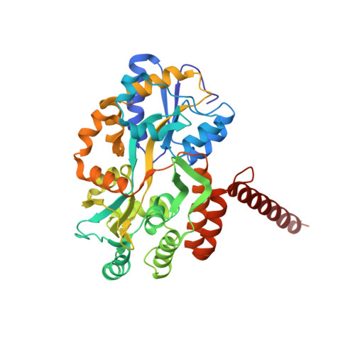
Mcm10 self-association is mediated by an N-terminal coiled-coil domain.

Du, W.Josephrajan, A.Adhikary, S.Bowles, T.Bielinsky, A.K.Eichman, B.F.

(2013) PLoS One 8: e70518-e70518

  • DOI: https://doi.org/10.1371/journal.pone.0070518
  • Primary Citation of Related Structures:  
    4JBZ

  • PubMed Abstract: 

    Minichromosome maintenance protein 10 (Mcm10) is an essential eukaryotic DNA-binding replication factor thought to serve as a scaffold to coordinate enzymatic activities within the replisome. Mcm10 appears to function as an oligomer rather than in its monomeric form (or rather than as a monomer). However, various orthologs have been found to contain 1, 2, 3, 4, or 6 subunits and thus, this issue has remained controversial. Here, we show that self-association of Xenopus laevis Mcm10 is mediated by a conserved coiled-coil (CC) motif within the N-terminal domain (NTD). Crystallographic analysis of the CC at 2.4 Å resolution revealed a three-helix bundle, consistent with the formation of both dimeric and trimeric Mcm10 CCs in solution. Mutation of the side chains at the subunit interface disrupted in vitro dimerization of both the CC and the NTD as monitored by analytical ultracentrifugation. In addition, the same mutations also impeded self-interaction of the full-length protein in vivo, as measured by yeast-two hybrid assays. We conclude that Mcm10 likely forms dimers or trimers to promote its diverse functions during DNA replication.


  • Organizational Affiliation

    Department of Biological Sciences, Vanderbilt University, Nashville, Tennessee, USA.


Macromolecules
Find similar proteins by:  (by identity cutoff)  |  3D Structure
Entity ID: 1
MoleculeChains Sequence LengthOrganismDetailsImage
MALTOSE-BINDING PERIPLASMIC PROTEIN FUSED WITH XENOPUS LAEVIS MCM10 COILED-COIL REGION
A, B, C
403Escherichia coli MS 21-1Xenopus laevis
This entity is chimeric
Mutation(s): 7 
Gene Names: HMPREF9530_03068Mcm10
UniProt
Find proteins for P0AEX9 (Escherichia coli (strain K12))
Explore P0AEX9 
Go to UniProtKB:  P0AEX9
Find proteins for Q5EAW4 (Xenopus laevis)
Explore Q5EAW4 
Go to UniProtKB:  Q5EAW4
Entity Groups  
Sequence Clusters30% Identity50% Identity70% Identity90% Identity95% Identity100% Identity
UniProt GroupsP0AEX9Q5EAW4
Sequence Annotations
Expand
  • Reference Sequence
Oligosaccharides

Help

Entity ID: 2
MoleculeChains Length2D Diagram Glycosylation3D Interactions
alpha-D-glucopyranose-(1-4)-alpha-D-glucopyranose
D, E, F
2N/A
Glycosylation Resources
GlyTouCan:  G07411ON
GlyCosmos:  G07411ON
Biologically Interesting Molecules (External Reference) 1 Unique
Experimental Data & Validation

Experimental Data

  • Method: X-RAY DIFFRACTION
  • Resolution: 2.40 Å
  • R-Value Free: 0.205 
  • R-Value Work: 0.164 
  • R-Value Observed: 0.166 
  • Space Group: C 1 2 1
Unit Cell:
Length ( Å )Angle ( ˚ )
a = 187.377α = 90
b = 116.618β = 103.48
c = 73.395γ = 90
Software Package:
Software NamePurpose
HKL-2000data collection
PHASERphasing
PHENIXrefinement
HKL-2000data reduction
HKL-2000data scaling

Structure Validation

View Full Validation Report


Entry History 

Deposition Data

Revision History  (Full details and data files)

  • Version 1.0: 2013-12-11
    Type: Initial release
  • Version 1.1: 2014-01-01
    Changes: Other
  • Version 1.2: 2017-08-09
    Changes: Refinement description, Source and taxonomy
  • Version 2.0: 2020-07-29
    Type: Remediation
    Reason: Carbohydrate remediation
    Changes: Atomic model, Data collection, Database references, Derived calculations, Non-polymer description, Structure summary
  • Version 2.1: 2023-09-20
    Changes: Data collection, Database references, Refinement description, Structure summary