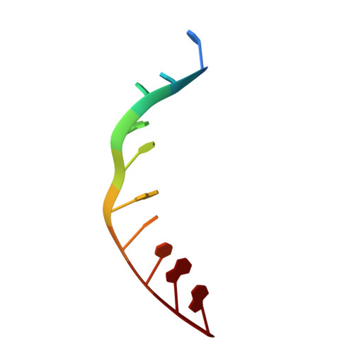
X-ray crystal structure of the ruthenium complex [Ru(phen)2(dppz)]2+ bound to d(TCGGTACCGA)
Niyazi, H., Teixeira, S., Mitchell, E., Forsyth, T., Cardin, C.To be published.
Experimental Data Snapshot
| Ligands 2 Unique | |||||
|---|---|---|---|---|---|
| ID | Chains | Name / Formula / InChI Key | 2D Diagram | 3D Interactions | |
| RKP Query on RKP | B [auth A], C [auth A] | Lambda-Ru(phen)2(dppz) complex C42 H26 N8 Ru OYSRBLHMGIHFCB-UHFFFAOYSA-N |  | ||
| BA Query on BA | D [auth A] | BARIUM ION Ba XDFCIPNJCBUZJN-UHFFFAOYSA-N |  | ||
| Length ( Å ) | Angle ( ˚ ) |
|---|---|
| a = 52.45 | α = 90 |
| b = 52.45 | β = 90 |
| c = 32.58 | γ = 90 |
| Software Name | Purpose |
|---|---|
| MxCuBE | data collection |
| SHELXS | phasing |
| REFMAC | refinement |
| MOSFLM | data reduction |
| SCALA | data scaling |
RCSB PDB (citation) is hosted by
RCSB PDB is a member of the