4N0Y

Structure of the Hepatitis C Envelope Glycoprotein E1 antigenic region 314-324 bound to the cross-neutralizing antibody IGH526


Experimental Data Snapshot

  • Method: X-RAY DIFFRACTION
  • Resolution: 1.75 Å
  • R-Value Free: 0.210 
  • R-Value Work: 0.179 
  • R-Value Observed: 0.180 

wwPDB Validation   3D Report Full Report


This is version 1.1 of the entry. See complete history


Literature

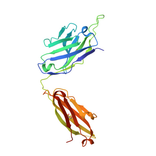
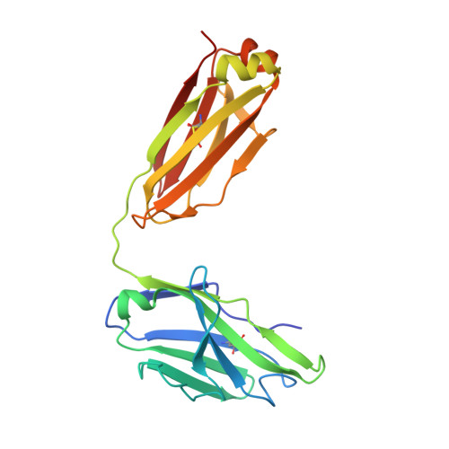
Structure of Hepatitis C Virus Envelope Glycoprotein E1 Antigenic Site 314-324 in Complex with Antibody IGH526.

Kong, L.Kadam, R.U.Giang, E.Ruwona, T.B.Nieusma, T.Culhane, J.C.Stanfield, R.L.Dawson, P.E.Wilson, I.A.Law, M.

(2015) J Mol Biol 427: 2617-2628

  • DOI: https://doi.org/10.1016/j.jmb.2015.06.012
  • Primary Citation of Related Structures:  
    4N0Y

  • PubMed Abstract: 

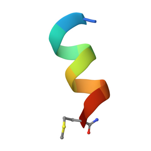
    Hepatitis C virus (HCV) is a positive-strand RNA virus within the Flaviviridae family. The viral "spike" of HCV is formed by two envelope glycoproteins, E1 and E2, which together mediate viral entry by engaging host receptors and undergoing conformational changes to facilitate membrane fusion. While E2 can be readily produced in the absence of E1, E1 cannot be expressed without E2 and few reagents, including monoclonal antibodies (mAbs), are available for study of this essential HCV glycoprotein. A human mAb to E1, IGH526, was previously reported to cross-neutralize different HCV isolates, and therefore, we sought to further characterize the IGH526 neutralizing epitope to obtain information for vaccine design. We found that mAb IGH526 bound to a discontinuous epitope, but with a major component corresponding to E1 residues 314-324. The crystal structure of IGH526 Fab with this E1 glycopeptide at 1.75Å resolution revealed that the antibody binds to one face of an α-helical peptide. Single mutations on the helix substantially lowered IGH526 binding but did not affect neutralization, indicating either that multiple mutations are required or that additional regions are recognized by the antibody in the context of the membrane-associated envelope oligomer. Molecular dynamics simulations indicate that the free peptide is flexible in solution, suggesting that it requires stabilization for use as a candidate vaccine immunogen.


  • Organizational Affiliation

    Department of Integrative Structural and Computational Biology, The Scripps Research Institute, La Jolla, CA 92037, USA.


Macromolecules
Find similar proteins by:  (by identity cutoff)  |  3D Structure
Entity ID: 1
MoleculeChains Sequence LengthOrganismDetailsImage
IGH526 Heavy ChainA [auth H]231Homo sapiensMutation(s): 0 
Entity Groups  
Sequence Clusters30% Identity50% Identity70% Identity90% Identity95% Identity100% Identity
Sequence Annotations
Expand
  • Reference Sequence
Find similar proteins by:  (by identity cutoff)  |  3D Structure
Entity ID: 2
MoleculeChains Sequence LengthOrganismDetailsImage
IGH526 Light ChainB [auth L]218Homo sapiensMutation(s): 0 
Entity Groups  
Sequence Clusters30% Identity50% Identity70% Identity90% Identity95% Identity100% Identity
Sequence Annotations
Expand
  • Reference Sequence

Find similar proteins by:  Sequence   |   3D Structure  

Entity ID: 3
MoleculeChains Sequence LengthOrganismDetailsImage
HCV E1 peptideC [auth A]12Hepacivirus hominisMutation(s): 1 
UniProt
Find proteins for P27958 (Hepatitis C virus genotype 1a (isolate H77))
Explore P27958 
Go to UniProtKB:  P27958
Entity Groups  
Sequence Clusters30% Identity50% Identity70% Identity90% Identity95% Identity100% Identity
UniProt GroupP27958
Sequence Annotations
Expand
  • Reference Sequence
Experimental Data & Validation

Experimental Data

  • Method: X-RAY DIFFRACTION
  • Resolution: 1.75 Å
  • R-Value Free: 0.210 
  • R-Value Work: 0.179 
  • R-Value Observed: 0.180 
  • Space Group: P 21 21 21
Unit Cell:
Length ( Å )Angle ( ˚ )
a = 66.326α = 90
b = 67.628β = 90
c = 120.719γ = 90
Software Package:
Software NamePurpose
Blu-Icedata collection
PHASERphasing
PHENIXrefinement
HKL-2000data reduction
SCALEPACKdata scaling

Structure Validation

View Full Validation Report


Entry History 

Deposition Data

Revision History  (Full details and data files)

  • Version 1.0: 2015-04-01
    Type: Initial release
  • Version 1.1: 2015-08-26
    Changes: Database references