


Mapping the binding surface of Cyclophilin A.
Mcnae, I.W., Dornan, D., Patterson, A.F., Wear, M.A., Blackburn, E.A., Walkinshaw, M.D.To be published.
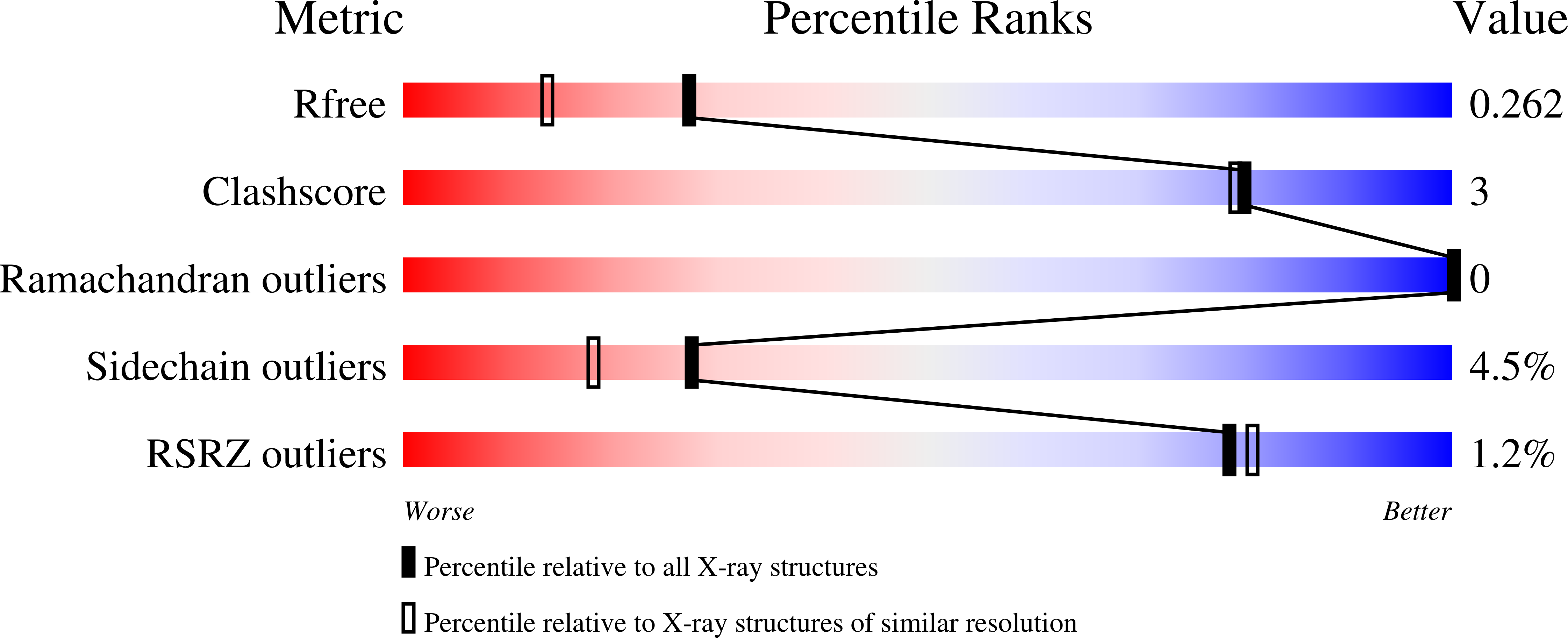
Experimental Data Snapshot
wwPDB Validation 3D Report Full Report
Entity ID: 1 | |||||
|---|---|---|---|---|---|
| Molecule | Chains | Sequence Length | Organism | Details | Image |
| Peptidyl-prolyl cis-trans isomerase A | 165 | Homo sapiens | Mutation(s): 0 Gene Names: PPIA, CYPA EC: 5.2.1.8 | 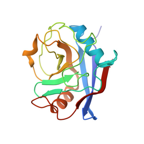 | |
UniProt & NIH Common Fund Data Resources | |||||
Find proteins for P62937 (Homo sapiens) Explore P62937 Go to UniProtKB: P62937 | |||||
PHAROS: P62937 GTEx: ENSG00000196262 | |||||
Entity Groups | |||||
| Sequence Clusters | 30% Identity50% Identity70% Identity90% Identity95% Identity100% Identity | ||||
| UniProt Group | P62937 | ||||
Sequence AnnotationsExpand | |||||
| |||||
| Ligands 1 Unique | |||||
|---|---|---|---|---|---|
| ID | Chains | Name / Formula / InChI Key | 2D Diagram | 3D Interactions | |
| WM1 Query on WM1 | B [auth A], C [auth A], D [auth A] | pyridine-2-carboxamide C6 H6 N2 O IBBMAWULFFBRKK-UHFFFAOYSA-N |  | ||
| Length ( Å ) | Angle ( ˚ ) |
|---|---|
| a = 42.81 | α = 90 |
| b = 54.28 | β = 90 |
| c = 87.14 | γ = 90 |
| Software Name | Purpose |
|---|---|
| MAR345dtb | data collection |
| PHASER | phasing |
| REFMAC | refinement |
| MOSFLM | data reduction |
| SCALA | data scaling |
RCSB PDB (citation) is hosted by
RCSB PDB is a member of the

