4OY3

Crystal Structure of the Helicobacter pylori MTAN-D198N mutant with S-Adenosylhomocysteine in the active site


Experimental Data Snapshot

  • Method: X-RAY DIFFRACTION
  • Resolution: 1.20 Å
  • R-Value Free: 0.145 
  • R-Value Work: 0.130 
  • R-Value Observed: 0.130 

wwPDB Validation   3D Report Full Report


Ligand Structure Quality Assessment 


This is version 1.1 of the entry. See complete history


Literature

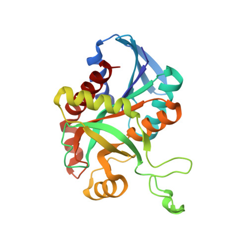
Crystal structures of the Helicobacter pylori MTAN enzyme reveal specific interactions between S-adenosylhomocysteine and the 5'-alkylthio binding subsite.

Mishra, V.Ronning, D.R.

(2012) Biochemistry 51: 9763-9772

  • DOI: https://doi.org/10.1021/bi301221k
  • Primary Citation of Related Structures:  
    4OJT, 4OY3, 4P54

  • PubMed Abstract: 

    The bacterial 5'-methylthioadenosine/S-adenosylhomocysteine nucleosidase (MTAN) enzyme is a multifunctional enzyme that catalyzes the hydrolysis of the N-ribosidic bond of at least four different adenosine-based metabolites: S-adenosylhomocysteine (SAH), 5'-methylthioadenosine (MTA), 5'-deoxyadenosine (5'-DOA), and 6-amino-6-deoxyfutalosine. These activities place the enzyme at the hub of seven fundamental bacterial metabolic pathways: S-adenosylmethionine (SAM) utilization, polyamine biosynthesis, the purine salvage pathway, the methionine salvage pathway, the SAM radical pathways, autoinducer-2 biosynthesis, and menaquinone biosynthesis. The last pathway makes MTAN essential for Helicobacter pylori viability. Although structures of various bacterial and plant MTANs have been described, the interactions between the homocysteine moiety of SAH and the 5'-alkylthiol binding site of MTAN have never been resolved. We have determined crystal structures of an inactive mutant form of H. pylori MTAN bound to MTA and SAH to 1.63 and 1.20 Å, respectively. The active form of MTAN was also crystallized in the presence of SAH, allowing the determination of the structure of a ternary enzyme-product complex resolved at 1.50 Å. These structures identify interactions between the homocysteine moiety and the 5'-alkylthiol binding site of the enzyme. This information can be leveraged for the development of species-specific MTAN inhibitors that prevent the growth of H. pylori.


  • Organizational Affiliation

    Department of Chemistry, University of Toledo, Toledo, OH 43606, USA.


Macromolecules
Find similar proteins by:  (by identity cutoff)  |  3D Structure
Entity ID: 1
MoleculeChains Sequence LengthOrganismDetailsImage
Aminodeoxyfutalosine nucleosidase231Helicobacter pylori J99Mutation(s): 1 
Gene Names: mtnNmtnjhp_0082
EC: 3.2.2.30 (PDB Primary Data), 3.2.2.9 (PDB Primary Data)
UniProt
Find proteins for Q9ZMY2 (Helicobacter pylori (strain J99 / ATCC 700824))
Explore Q9ZMY2 
Go to UniProtKB:  Q9ZMY2
Entity Groups  
Sequence Clusters30% Identity50% Identity70% Identity90% Identity95% Identity100% Identity
UniProt GroupQ9ZMY2
Sequence Annotations
Expand
  • Reference Sequence
Small Molecules
Ligands 2 Unique
IDChains Name / Formula / InChI Key2D Diagram3D Interactions
SAH
Query on SAH

Download Ideal Coordinates CCD File 
B [auth A]S-ADENOSYL-L-HOMOCYSTEINE
C14 H20 N6 O5 S
ZJUKTBDSGOFHSH-WFMPWKQPSA-N
CL
Query on CL

Download Ideal Coordinates CCD File 
C [auth A]CHLORIDE ION
Cl
VEXZGXHMUGYJMC-UHFFFAOYSA-M
Experimental Data & Validation

Experimental Data

  • Method: X-RAY DIFFRACTION
  • Resolution: 1.20 Å
  • R-Value Free: 0.145 
  • R-Value Work: 0.130 
  • R-Value Observed: 0.130 
  • Space Group: P 32 2 1
Unit Cell:
Length ( Å )Angle ( ˚ )
a = 80.733α = 90
b = 80.733β = 90
c = 67.199γ = 120
Software Package:
Software NamePurpose
PHENIXrefinement

Structure Validation

View Full Validation Report


Ligand Structure Quality Assessment 


Entry History 

Deposition Data

Revision History  (Full details and data files)

  • Version 1.0: 2014-12-17
    Type: Initial release
  • Version 1.1: 2023-12-27
    Changes: Data collection, Database references, Derived calculations, Source and taxonomy