4UBS

The crystal structure of cytochrome P450 105D7 from Streptomyces avermitilis in complex with Diclofenac


Experimental Data Snapshot

  • Method: X-RAY DIFFRACTION
  • Resolution: 2.20 Å
  • R-Value Free: 0.228 
  • R-Value Work: 0.177 
  • R-Value Observed: 0.179 

wwPDB Validation   3D Report Full Report


Ligand Structure Quality Assessment 


This is version 1.3 of the entry. See complete history


Literature

Structural basis for the 4'-hydroxylation of diclofenac by a microbial cytochrome P450 monooxygenase.

Xu, L.H.Ikeda, H.Liu, L.Arakawa, T.Wakagi, T.Shoun, H.Fushinobu, S.

(2015) Appl Microbiol Biotechnol 99: 3081-3091

  • DOI: https://doi.org/10.1007/s00253-014-6148-y
  • Primary Citation of Related Structures:  
    4UBS

  • PubMed Abstract: 

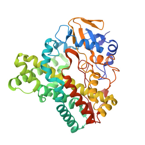
    Diclofenac is a nonsteroidal anti-inflammatory drug. It undergoes hydroxylation by mammalian cytochrome P450 enzymes at 4'- and/or 5'-positions. A bacterial P450 enzyme, CYP105D7 from Streptomyces avermitilis, has been shown to catalyze hydroxylation of 1-deoxypentalenic acid and an isoflavone daidzein. Here, we demonstrated that CYP105D7 also catalyzes hydroxylation of diclofenac at the C4'-position. A spectroscopic analysis showed that CYP105D7 binds diclofenac in a slightly cooperative manner with an affinity of 65 μM and a Hill coefficient of 1.16. The crystal structure of CYP105D7 in complex with diclofenac was determined at 2.2 Å resolution. The distal pocket of CYP105D7 contains two diclofenac molecules, illustrating drug recognition with a double-ligand-binding mode. The C3' and C4' atoms of the dichlorophenyl ring of one diclofenac molecule are positioned near the heme iron, suggesting that it is positioned appropriately for aromatic hydroxylation to yield the 4'-hydroxylated product. However, recognition of diclofenac by CYP105D7 was completely different from that of rabbit CYP2C5, which binds one diclofenac molecule with a cluster of water molecules. The distal pocket of CYP105D7 contains four arginine residues, forming a wall of the substrate-binding pocket, and the arginine residues are conserved in bacterial P450s in the CYP105 family.


  • Organizational Affiliation

    Ocean College, Zhejiang University, 866 Yuhangtang Road, Hangzhou, Zhejiang, 310058, China, lianhuaxu@zju.edu.cn.


Macromolecules
Find similar proteins by:  (by identity cutoff)  |  3D Structure
Entity ID: 1
MoleculeChains Sequence LengthOrganismDetailsImage
Pentalenic acid synthase393Streptomyces avermitilis MA-4680 = NBRC 14893Mutation(s): 0 
Gene Names: cyp105d7cyp28SAV_7469
EC: 1.14.15.11
UniProt
Find proteins for Q825I8 (Streptomyces avermitilis (strain ATCC 31267 / DSM 46492 / JCM 5070 / NBRC 14893 / NCIMB 12804 / NRRL 8165 / MA-4680))
Explore Q825I8 
Go to UniProtKB:  Q825I8
Entity Groups  
Sequence Clusters30% Identity50% Identity70% Identity90% Identity95% Identity100% Identity
UniProt GroupQ825I8
Sequence Annotations
Expand
  • Reference Sequence
Small Molecules
Ligands 5 Unique
IDChains Name / Formula / InChI Key2D Diagram3D Interactions
HEM
Query on HEM

Download Ideal Coordinates CCD File 
B [auth A]PROTOPORPHYRIN IX CONTAINING FE
C34 H32 Fe N4 O4
KABFMIBPWCXCRK-RGGAHWMASA-L
DIF
Query on DIF

Download Ideal Coordinates CCD File 
C [auth A],
D [auth A]
2-[2,6-DICHLOROPHENYL)AMINO]BENZENEACETIC ACID
C14 H11 Cl2 N O2
DCOPUUMXTXDBNB-UHFFFAOYSA-N
PGE
Query on PGE

Download Ideal Coordinates CCD File 
E [auth A],
F [auth A],
G [auth A],
H [auth A],
I [auth A]
TRIETHYLENE GLYCOL
C6 H14 O4
ZIBGPFATKBEMQZ-UHFFFAOYSA-N
PEG
Query on PEG

Download Ideal Coordinates CCD File 
J [auth A]
K [auth A]
L [auth A]
M [auth A]
N [auth A]
J [auth A],
K [auth A],
L [auth A],
M [auth A],
N [auth A],
O [auth A],
P [auth A],
Q [auth A],
R [auth A],
S [auth A],
T [auth A],
U [auth A],
V [auth A],
W [auth A]
DI(HYDROXYETHYL)ETHER
C4 H10 O3
MTHSVFCYNBDYFN-UHFFFAOYSA-N
PO4
Query on PO4

Download Ideal Coordinates CCD File 
X [auth A]PHOSPHATE ION
O4 P
NBIIXXVUZAFLBC-UHFFFAOYSA-K
Binding Affinity Annotations 
IDSourceBinding Affinity
DIF Binding MOAD:  4UBS Kd: 6.50e+4 (nM) from 1 assay(s)
Experimental Data & Validation

Experimental Data

  • Method: X-RAY DIFFRACTION
  • Resolution: 2.20 Å
  • R-Value Free: 0.228 
  • R-Value Work: 0.177 
  • R-Value Observed: 0.179 
  • Space Group: P 31 2 1
Unit Cell:
Length ( Å )Angle ( ˚ )
a = 139.897α = 90
b = 139.897β = 90
c = 65.268γ = 120
Software Package:
Software NamePurpose
REFMACrefinement
PDB_EXTRACTdata extraction
ADSCdata collection
MOLREPmodel building
HKLdata reduction

Structure Validation

View Full Validation Report


Ligand Structure Quality Assessment 


Entry History & Funding Information

Deposition Data


Funding OrganizationLocationGrant Number
the fundamental research funds for the central universitiesChina2013QNA4042

Revision History  (Full details and data files)

  • Version 1.0: 2014-11-05
    Type: Initial release
  • Version 1.1: 2015-04-01
    Changes: Database references
  • Version 1.2: 2020-01-29
    Changes: Data collection, Database references, Derived calculations, Other, Source and taxonomy
  • Version 1.3: 2024-03-20
    Changes: Data collection, Database references, Refinement description