4UP4

Structure of the recombinant lectin PVL from Psathyrella velutina in complex with GlcNAcb-D-1,3Galactoside


Experimental Data Snapshot

  • Method: X-RAY DIFFRACTION
  • Resolution: 1.95 Å
  • R-Value Free: 0.237 
  • R-Value Work: 0.190 
  • R-Value Observed: 0.192 

wwPDB Validation   3D Report Full Report


Ligand Structure Quality Assessment 


This is version 2.1 of the entry. See complete history


Literature

A Recombinant Fungal Lectin for Labeling Truncated Glycans on Human Cancer Cells.

Audfray, A.Beldjoudi, M.Breiman, A.Hurbin, A.Boos, I.Unverzagt, C.Bouras, M.Lantuejoul, S.Coll, J.Varrot, A.Le Pendu, J.Busser, B.Imberty, A.

(2015) PLoS One 10: 01281

  • DOI: https://doi.org/10.1371/journal.pone.0128190
  • Primary Citation of Related Structures:  
    4UP4

  • PubMed Abstract: 

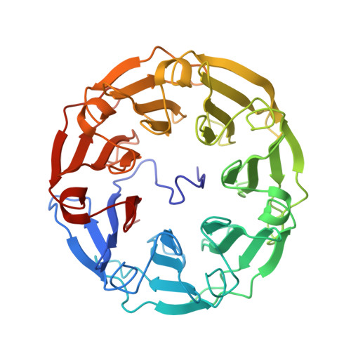
    Cell surface glycoconjugates present alterations of their structures in chronic diseases and distinct oligosaccharide epitopes have been associated with cancer. Among them, truncated glycans present terminal non-reducing β-N-acetylglucosamine (GlcNAc) residues that are rare on healthy tissues. Lectins from unconventional sources such as fungi or algi provide novel markers that bind specifically to such epitopes, but their availability may be challenging. A GlcNAc-binding lectin from the fruiting body of the fungus Psathyrella velutina (PVL) has been produced in good yield in bacterial culture. A strong specificity for terminal GlcNAc residues was evidenced by glycan array. Affinity values obtained by microcalorimetry and surface plasmon resonance demonstrated a micromolar affinity for GlcNAcβ1-3Gal epitopes and for biantennary N-glycans with GlcNAcβ1-2Man capped branches. Crystal structure of PVL complexed with GlcNAcβ1-3Gal established the structural basis of the specificity. Labeling of several types of cancer cells and use of inhibitors of glycan metabolism indicated that rPVL binds to terminal GlcNAc but also to sialic acid (Neu5Ac). Analysis of glycosyltransferase expression confirmed the higher amount of GlcNAc present on cancer cells. rPVL binding is specific to cancer tissue and weak or no labeling is observed for healthy ones, except for stomach glands that present unique αGlcNAc-presenting mucins. In lung, breast and colon carcinomas, a clear delineation could be observed between cancer regions and surrounding healthy tissues. PVL is therefore a useful tool for labeling agalacto-glycans in cancer or other diseases.


  • Organizational Affiliation

    CERMAV, UPR5301, CNRS, University Grenoble Alpes, 38041 Grenoble, France.


Macromolecules
Find similar proteins by:  (by identity cutoff)  |  3D Structure
Entity ID: 1
MoleculeChains Sequence LengthOrganismDetailsImage
LECTIN PVL
A, B
401Lacrymaria velutinaMutation(s): 0 
UniProt
Find proteins for Q309D1 (Lacrymaria velutina)
Explore Q309D1 
Go to UniProtKB:  Q309D1
Entity Groups  
Sequence Clusters30% Identity50% Identity70% Identity90% Identity95% Identity100% Identity
UniProt GroupQ309D1
Sequence Annotations
Expand
  • Reference Sequence
Oligosaccharides

Help

Entity ID: 2
MoleculeChains Length2D Diagram Glycosylation3D Interactions
2-acetamido-2-deoxy-beta-D-glucopyranose-(1-3)-beta-D-galactopyranose
C, D, E, F, G
C, D, E, F, G, H, I, J, K, L
2N/A
Glycosylation Resources
GlyTouCan:  G33476GF
GlyCosmos:  G33476GF
GlyGen:  G33476GF
Small Molecules
Ligands 3 Unique
IDChains Name / Formula / InChI Key2D Diagram3D Interactions
NDG
Query on NDG

Download Ideal Coordinates CCD File 
P [auth A],
U [auth B]
2-acetamido-2-deoxy-alpha-D-glucopyranose
C8 H15 N O6
OVRNDRQMDRJTHS-PVFLNQBWSA-N
NAG
Query on NAG

Download Ideal Coordinates CCD File 
Q [auth A]2-acetamido-2-deoxy-beta-D-glucopyranose
C8 H15 N O6
OVRNDRQMDRJTHS-FMDGEEDCSA-N
CA
Query on CA

Download Ideal Coordinates CCD File 
M [auth A]
N [auth A]
O [auth A]
R [auth B]
S [auth B]
M [auth A],
N [auth A],
O [auth A],
R [auth B],
S [auth B],
T [auth B]
CALCIUM ION
Ca
BHPQYMZQTOCNFJ-UHFFFAOYSA-N
Experimental Data & Validation

Experimental Data

  • Method: X-RAY DIFFRACTION
  • Resolution: 1.95 Å
  • R-Value Free: 0.237 
  • R-Value Work: 0.190 
  • R-Value Observed: 0.192 
  • Space Group: P 21 21 21
Unit Cell:
Length ( Å )Angle ( ˚ )
a = 54.814α = 90
b = 97.468β = 90
c = 158.65γ = 90
Software Package:
Software NamePurpose
REFMACrefinement
XDSdata reduction
Aimlessdata scaling
PHASERphasing

Structure Validation

View Full Validation Report


Ligand Structure Quality Assessment 


Entry History 

Deposition Data

Revision History  (Full details and data files)

  • Version 1.0: 2015-01-28
    Type: Initial release
  • Version 1.1: 2015-06-17
    Changes: Database references
  • Version 1.2: 2017-05-03
    Changes: Database references
  • Version 2.0: 2020-07-29
    Type: Remediation
    Reason: Carbohydrate remediation
    Changes: Atomic model, Data collection, Derived calculations, Other, Structure summary
  • Version 2.1: 2024-01-10
    Changes: Data collection, Database references, Derived calculations, Refinement description, Structure summary