4V0U

The crystal structure of ternary PP1G-PPP1R15B and G-actin complex


Experimental Data Snapshot

  • Method: X-RAY DIFFRACTION
  • Resolution: 7.88 Å
  • R-Value Free: 0.400 
  • R-Value Work: 0.370 
  • R-Value Observed: 0.372 

wwPDB Validation   3D Report Full Report


Ligand Structure Quality Assessment 


This is version 1.2 of the entry. See complete history


Literature

G-actin provides substrate-specificity to eukaryotic initiation factor 2 alpha holophosphatases.

Chen, R.Rato, C.Yan, Y.Crespillo-Casado, A.Clarke, H.J.Harding, H.P.Marciniak, S.J.Read, R.J.Ron, D.

(2015) Elife 4

  • DOI: https://doi.org/10.7554/eLife.04871
  • Primary Citation of Related Structures:  
    4V0U, 4V0V, 4V0W, 4V0X

  • PubMed Abstract: 

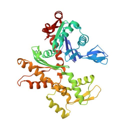
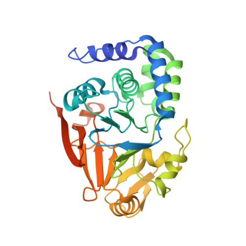
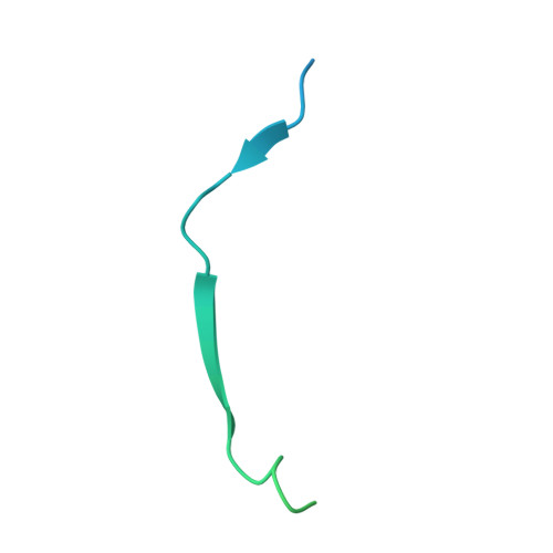
    Dephosphorylation of eukaryotic translation initiation factor 2a (eIF2a) restores protein synthesis at the waning of stress responses and requires a PP1 catalytic subunit and a regulatory subunit, PPP1R15A/GADD34 or PPP1R15B/CReP. Surprisingly, PPP1R15-PP1 binary complexes reconstituted in vitro lacked substrate selectivity. However, selectivity was restored by crude cell lysate or purified G-actin, which joined PPP1R15-PP1 to form a stable ternary complex. In crystal structures of the non-selective PPP1R15B-PP1G complex, the functional core of PPP1R15 made multiple surface contacts with PP1G, but at a distance from the active site, whereas in the substrate-selective ternary complex, actin contributes to one face of a platform encompassing the active site. Computational docking of the N-terminal lobe of eIF2a at this platform placed phosphorylated serine 51 near the active site. Mutagenesis of predicted surface-contacting residues enfeebled dephosphorylation, suggesting that avidity for the substrate plays an important role in imparting specificity on the PPP1R15B-PP1G-actin ternary complex.


  • Organizational Affiliation

    Cambridge Institute for Medical Research, University of Cambridge, Cambridge, United Kingdom.


Macromolecules
Find similar proteins by:  (by identity cutoff)  |  3D Structure
Entity ID: 1
MoleculeChains Sequence LengthOrganismDetailsImage
ACTIN, ALPHA SKELETAL MUSCLE
A, B, C, L, M
375Oryctolagus cuniculusMutation(s): 0 
UniProt
Find proteins for P68135 (Oryctolagus cuniculus)
Explore P68135 
Go to UniProtKB:  P68135
Entity Groups  
Sequence Clusters30% Identity50% Identity70% Identity90% Identity95% Identity100% Identity
UniProt GroupP68135
Sequence Annotations
Expand
  • Reference Sequence
Find similar proteins by:  (by identity cutoff)  |  3D Structure
Entity ID: 2
MoleculeChains Sequence LengthOrganismDetailsImage
SERINE/THREONINE-PROTEIN PHOSPHATASE PP1-GAMMA CATALYTIC SUBUNIT
D, F, H, J, N
323Mus musculusMutation(s): 0 
EC: 3.1.3.16
UniProt & NIH Common Fund Data Resources
Find proteins for P63087 (Mus musculus)
Explore P63087 
Go to UniProtKB:  P63087
IMPC:  MGI:104872
Entity Groups  
Sequence Clusters30% Identity50% Identity70% Identity90% Identity95% Identity100% Identity
UniProt GroupP63087
Sequence Annotations
Expand
  • Reference Sequence
Find similar proteins by:  (by identity cutoff)  |  3D Structure
Entity ID: 3
MoleculeChains Sequence LengthOrganismDetailsImage
PROTEIN PHOSPHATASE 1 REGULATORY SUBUNIT 15B
E, G, I, K, O
84Homo sapiensMutation(s): 0 
UniProt & NIH Common Fund Data Resources
Find proteins for Q5SWA1 (Homo sapiens)
Explore Q5SWA1 
Go to UniProtKB:  Q5SWA1
PHAROS:  Q5SWA1
GTEx:  ENSG00000158615 
Entity Groups  
Sequence Clusters30% Identity50% Identity70% Identity90% Identity95% Identity100% Identity
UniProt GroupQ5SWA1
Sequence Annotations
Expand
  • Reference Sequence
Small Molecules
Ligands 3 Unique
IDChains Name / Formula / InChI Key2D Diagram3D Interactions
ATP
Query on ATP

Download Ideal Coordinates CCD File 
EA [auth L],
GA [auth M],
Q [auth A],
S [auth B],
U [auth C]
ADENOSINE-5'-TRIPHOSPHATE
C10 H16 N5 O13 P3
ZKHQWZAMYRWXGA-KQYNXXCUSA-N
LAB
Query on LAB

Download Ideal Coordinates CCD File 
DA [auth L],
FA [auth M],
P [auth A],
R [auth B],
T [auth C]
LATRUNCULIN B
C20 H29 N O5 S
NSHPHXHGRHSMIK-JRIKCGFMSA-N
MN
Query on MN

Download Ideal Coordinates CCD File 
AA [auth H]
BA [auth J]
CA [auth J]
HA [auth N]
IA [auth N]
AA [auth H],
BA [auth J],
CA [auth J],
HA [auth N],
IA [auth N],
V [auth D],
W [auth D],
X [auth F],
Y [auth F],
Z [auth H]
MANGANESE (II) ION
Mn
WAEMQWOKJMHJLA-UHFFFAOYSA-N
Experimental Data & Validation

Experimental Data

  • Method: X-RAY DIFFRACTION
  • Resolution: 7.88 Å
  • R-Value Free: 0.400 
  • R-Value Work: 0.370 
  • R-Value Observed: 0.372 
  • Space Group: C 1 2 1
Unit Cell:
Length ( Å )Angle ( ˚ )
a = 103.91α = 90
b = 149.93β = 91.03
c = 318.72γ = 90
Software Package:
Software NamePurpose
REFMACrefinement
XDSdata reduction
Aimlessdata scaling
CCP4Iphasing

Structure Validation

View Full Validation Report


Ligand Structure Quality Assessment 


Entry History 

Deposition Data

Revision History  (Full details and data files)

  • Version 1.0: 2015-03-25
    Type: Initial release
  • Version 1.1: 2019-02-27
    Changes: Data collection, Database references, Other
  • Version 1.2: 2024-01-10
    Changes: Data collection, Database references, Derived calculations, Other, Refinement description