Human 3 alpha-hydroxysteroid dehydrogenase type 3: structural clues of 5 alpha-DHT reverse binding and enzyme down-regulation decreasing MCF7 cell growth.
Zhang, B., Hu, X.J., Wang, X.Q., Theriault, J.F., Zhu, D.W., Shang, P., Labrie, F., Lin, S.X.(2016) Biochem J 473: 1037-1046
- PubMed: 26929402
- DOI: https://doi.org/10.1042/BCJ20160083
- Primary Citation of Related Structures:
4XO6, 4XO7 - PubMed Abstract:
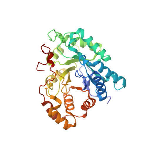
Human 3α-HSD3 (3α-hydroxysteroid dehydrogenase type 3) plays an essential role in the inactivation of the most potent androgen 5α-DHT (5α-dihydrotestosterone). The present study attempts to obtain the important structure of 3α-HSD3 in complex with 5α-DHT and to investigate the role of 3α-HSD3 in breast cancer cells. We report the crystal structure of human 3α-HSD3·NADP(+)·A-dione (5α-androstane-3,17-dione)/epi-ADT (epiandrosterone) complex, which was obtained by co-crystallization with 5α-DHT in the presence of NADP(+) Although 5α-DHT was introduced during the crystallization, oxidoreduction of 5α-DHT occurred. The locations of A-dione and epi-ADT were identified in the steroid-binding sites of two 3α-HSD3 molecules per crystal asymmetric unit. An overlay showed that A-dione and epi-ADT were oriented upside-down and flipped relative to each other, providing structural clues for 5α-DHT reverse binding in the enzyme with the generation of different products. Moreover, we report the crystal structure of the 3α-HSD3·NADP(+)·4-dione (4-androstene-3,17-dione) complex. When a specific siRNA (100 nM) was used to suppress 3α-HSD3 expression without interfering with 3α-HSD4, which shares a highly homologous active site, the 5α-DHT concentration increased, whereas MCF7 cell growth was suppressed. The present study provides structural clues for 5α-DHT reverse binding within 3α-HSD3, and demonstrates for the first time that down-regulation of 3α-HSD3 decreases MCF7 breast cancer cell growth.
Organizational Affiliation:
Laboratory of Molecular Endocrinology and Oncology, Centre Hospitalier Universitaire de Quebec Research Center (CHUL, CHU) and Laval University, Québec City, Québec, G1V4G2, Canada Key Laboratory for Space Bioscience and Biotechnology, Institute of Special Environmental Biophysics, School of Life Sciences, Northwestern Polytechnical University, Xi'an 710072, P.R. China.