5C8I

Joint X-ray/neutron structure of Human Carbonic Anhydrase II in complex with Methazolamide


Experimental Data Snapshot

  • Method: X-RAY DIFFRACTION
  • Resolution: 1.56 Å
  • R-Value Free: 0.221 
  • R-Value Work: 0.204 

  • Method: NEUTRON DIFFRACTION
  • Resolution: 2.20 Å
  • R-Value Free: 0.276 
  • R-Value Work: 0.225 

wwPDB Validation   3D Report Full Report


This is version 1.5 of the entry. See complete history


Literature

Neutron structure of human carbonic anhydrase II in complex with methazolamide: mapping the solvent and hydrogen-bonding patterns of an effective clinical drug.

Aggarwal, M.Kovalevsky, A.Y.Velazquez, H.Fisher, S.Z.Smith, J.C.McKenna, R.

(2016) IUCrJ 3: 319-325

  • DOI: https://doi.org/10.1107/S2052252516010514
  • Primary Citation of Related Structures:  
    5C8I

  • PubMed Abstract: 

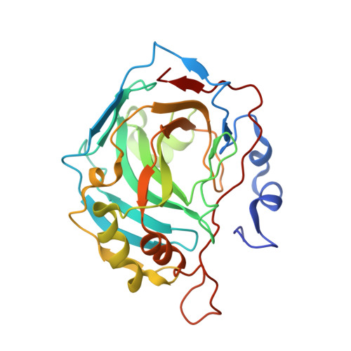
    Carbonic anhydrases (CAs; EC 4.2.1.1) catalyze the interconversion of CO 2 and HCO 3 - , and their inhibitors have long been used as diuretics and as a therapeutic treatment for many disorders such as glaucoma and epilepsy. Acetazolamide (AZM) and methazolamide (MZM, a methyl derivative of AZM) are two of the classical CA inhibitory drugs that have been used clinically for decades. The jointly refined X-ray/neutron structure of MZM in complex with human CA isoform II (hCA II) has been determined to a resolution of 2.2 Å with an R cryst of ∼16.0%. Presented in this article, along with only the second neutron structure of a clinical drug-bound hCA, is an in-depth structural comparison and analyses of differences in hydrogen-bonding network, water-molecule orientation and solvent displacement that take place upon the binding of AZM and MZM in the active site of hCA II. Even though MZM is slightly more hydrophobic and displaces more waters than AZM, the overall binding affinity ( K i ) for both of the drugs against hCA II is similar (∼10 n M ). The plausible reasons behind this finding have also been discussed using molecular dynamics and X-ray crystal structures of hCA II-MZM determined at cryotemperature and room temperature. This study not only allows a direct comparison of the hydrogen bonding, protonation states and solvent orientation/displacement of AZM and MZM, but also shows the significant effect that the methyl derivative has on the solvent organization in the hCA II active site.


  • Organizational Affiliation

    Biology and Soft Matter Division, Oak Ridge National Laboratory, Oak Ridge, TN 37831, USA.


Macromolecules
Find similar proteins by:  (by identity cutoff)  |  3D Structure
Entity ID: 1
MoleculeChains Sequence LengthOrganismDetailsImage
Carbonic anhydrase 2260Homo sapiensMutation(s): 0 
Gene Names: CA2
EC: 4.2.1.1
UniProt & NIH Common Fund Data Resources
Find proteins for P00918 (Homo sapiens)
Explore P00918 
Go to UniProtKB:  P00918
PHAROS:  P00918
GTEx:  ENSG00000104267 
Entity Groups  
Sequence Clusters30% Identity50% Identity70% Identity90% Identity95% Identity100% Identity
UniProt GroupP00918
Sequence Annotations
Expand
  • Reference Sequence
Small Molecules
Ligands 2 Unique
IDChains Name / Formula / InChI Key2D Diagram3D Interactions
MZM
Query on MZM

Download Ideal Coordinates CCD File 
C [auth A]N-(3-methyl-5-sulfamoyl-1,3,4-thiadiazol-2(3H)-ylidene)acetamide
C5 H8 N4 O3 S2
FLOSMHQXBMRNHR-DAXSKMNVSA-N
ZN
Query on ZN

Download Ideal Coordinates CCD File 
B [auth A]ZINC ION
Zn
PTFCDOFLOPIGGS-UHFFFAOYSA-N
Binding Affinity Annotations 
IDSourceBinding Affinity
MZM BindingDB:  5C8I Ki: min: 9, max: 1.40e+4 (nM) from 4 assay(s)
IC50: min: 4.8, max: 21 (nM) from 4 assay(s)
EC50: 40 (nM) from 1 assay(s)
Experimental Data & Validation

Experimental Data

  • Method: X-RAY DIFFRACTION
  • Resolution: 1.56 Å
  • R-Value Free: 0.221 
  • R-Value Work: 0.204 
  • Space Group: P 1 21 1
  • Method: NEUTRON DIFFRACTION
  • Resolution: 2.20 Å
  • R-Value Free: 0.276 
  • R-Value Work: 0.225 
  • Space Group: P 1 21 1
Unit Cell:
Length ( Å )Angle ( ˚ )
a = 42.893α = 90
b = 41.763β = 104.59
c = 72.949γ = 90
Software Package:
Software NamePurpose
nCNSrefinement
CNSphasing

Structure Validation

View Full Validation Report


Entry History & Funding Information

Deposition Data


Funding OrganizationLocationGrant Number
National Science Foundation (NSF, United States)United States0922719
Oak Ridge National Lab/Shull FellowshipUnited StatesNDSB0001

Revision History  (Full details and data files)

  • Version 1.0: 2016-07-20
    Type: Initial release
  • Version 1.1: 2016-09-07
    Changes: Database references
  • Version 1.2: 2017-09-20
    Changes: Advisory, Author supporting evidence, Derived calculations
  • Version 1.3: 2018-04-18
    Changes: Data collection, Database references
  • Version 1.4: 2019-11-27
    Changes: Author supporting evidence
  • Version 1.5: 2024-03-06
    Changes: Advisory, Data collection, Database references