5EA0

Structure of the antibody 7968 with human complement factor H-derived peptide


Experimental Data Snapshot

  • Method: X-RAY DIFFRACTION
  • Resolution: 2.00 Å
  • R-Value Free: 0.275 
  • R-Value Work: 0.196 
  • R-Value Observed: 0.202 

wwPDB Validation   3D Report Full Report


This is version 1.3 of the entry. See complete history


Literature

A Therapeutic Antibody for Cancer, Derived from Single Human B Cells.

Bushey, R.T.Moody, M.A.Nicely, N.L.Haynes, B.F.Alam, S.M.Keir, S.T.Bentley, R.C.Roy Choudhury, K.Gottlin, E.B.Campa, M.J.Liao, H.X.Patz, E.F.

(2016) Cell Rep 15: 1505-1513

  • DOI: https://doi.org/10.1016/j.celrep.2016.04.038
  • Primary Citation of Related Structures:  
    5EA0

  • PubMed Abstract: 

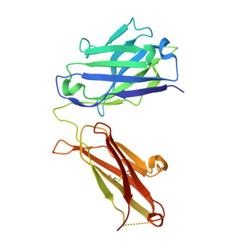
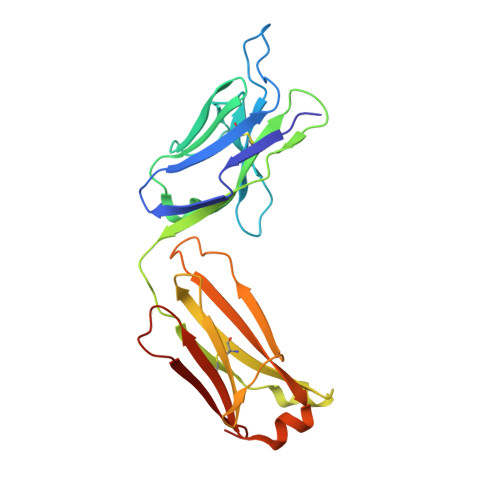
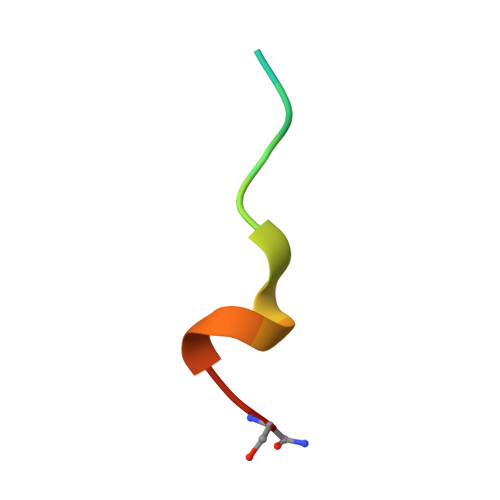
    Some patients with cancer never develop metastasis, and their host response might provide cues for innovative treatment strategies. We previously reported an association between autoantibodies against complement factor H (CFH) and early-stage lung cancer. CFH prevents complement-mediated cytotoxicity (CDC) by inhibiting formation of cell-lytic membrane attack complexes on self-surfaces. In an effort to translate these findings into a biologic therapy for cancer, we isolated and expressed DNA sequences encoding high-affinity human CFH antibodies directly from single, sorted B cells obtained from patients with the antibody. The co-crystal structure of a CFH antibody-target complex shows a conformational change in the target relative to the native structure. This recombinant CFH antibody causes complement activation and release of anaphylatoxins, promotes CDC of tumor cell lines, and inhibits tumor growth in vivo. The isolation of anti-tumor antibodies derived from single human B cells represents an alternative paradigm in antibody drug discovery.


  • Organizational Affiliation

    Department of Radiology, Duke University Medical Center, Durham, NC 27710, USA.


Macromolecules
Find similar proteins by:  (by identity cutoff)  |  3D Structure
Entity ID: 1
MoleculeChains Sequence LengthOrganismDetailsImage
Heavy chain of antibody 7968 Fab fragmentA [auth H]224Homo sapiensMutation(s): 0 
Entity Groups  
Sequence Clusters30% Identity50% Identity70% Identity90% Identity95% Identity100% Identity
Sequence Annotations
Expand
  • Reference Sequence
Find similar proteins by:  (by identity cutoff)  |  3D Structure
Entity ID: 2
MoleculeChains Sequence LengthOrganismDetailsImage
Light chain of antibody 7968 Fab fragmentB [auth L]220Homo sapiensMutation(s): 0 
Entity Groups  
Sequence Clusters30% Identity50% Identity70% Identity90% Identity95% Identity100% Identity
Sequence Annotations
Expand
  • Reference Sequence

Find similar proteins by:  Sequence   |   3D Structure  

Entity ID: 3
MoleculeChains Sequence LengthOrganismDetailsImage
Complement factor H-related protein 2C [auth P]15Homo sapiensMutation(s): 2 
UniProt & NIH Common Fund Data Resources
Find proteins for P36980 (Homo sapiens)
Explore P36980 
Go to UniProtKB:  P36980
PHAROS:  P36980
GTEx:  ENSG00000080910 
Entity Groups  
Sequence Clusters30% Identity50% Identity70% Identity90% Identity95% Identity100% Identity
UniProt GroupP36980
Sequence Annotations
Expand
  • Reference Sequence
Experimental Data & Validation

Experimental Data

  • Method: X-RAY DIFFRACTION
  • Resolution: 2.00 Å
  • R-Value Free: 0.275 
  • R-Value Work: 0.196 
  • R-Value Observed: 0.202 
  • Space Group: P 1 21 1
Unit Cell:
Length ( Å )Angle ( ˚ )
a = 51.28α = 90
b = 65.547β = 106.37
c = 71.267γ = 90
Software Package:
Software NamePurpose
PHENIXrefinement
HKL-2000data reduction
HKL-2000data scaling
PHENIXphasing

Structure Validation

View Full Validation Report


Entry History 

Deposition Data

Revision History  (Full details and data files)

  • Version 1.0: 2016-05-25
    Type: Initial release
  • Version 1.1: 2016-06-01
    Changes: Database references
  • Version 1.2: 2021-03-24
    Changes: Derived calculations, Source and taxonomy
  • Version 1.3: 2023-09-27
    Changes: Data collection, Database references, Refinement description