5G0N

Structure of rat neuronal nitric oxide synthase D597N mutant heme domain in complex with N1-(5-(2-(6-AMINO-4-METHYLPYRIDIN-2-YL)ETHYL) PYRIDIN-3-YL)-N1,N2-DIMETHYLETHANE-1,2-DIAMINE


Experimental Data Snapshot

  • Method: X-RAY DIFFRACTION
  • Resolution: 1.94 Å
  • R-Value Free: 0.216 
  • R-Value Work: 0.175 
  • R-Value Observed: 0.177 

wwPDB Validation   3D Report Full Report


Ligand Structure Quality Assessment 


This is version 1.2 of the entry. See complete history


Literature

Electrostatic Control of Isoform Selective Inhibitor Binding in Nitric Oxide Synthase.

Li, H.Wang, H.Kang, S.Silverman, R.B.Poulos, T.L.

(2016) Biochemistry 55: 3702

  • DOI: https://doi.org/10.1021/acs.biochem.6b00261
  • Primary Citation of Related Structures:  
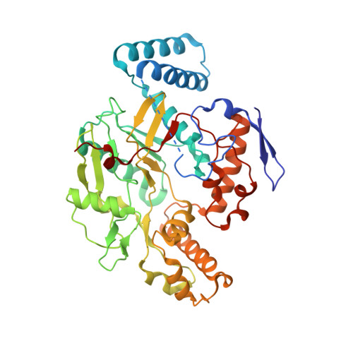
    5G0N, 5G0O, 5G0P

  • PubMed Abstract: 

    Development of potent and isoform selective nitric oxide synthase (NOS) inhibitors is challenging because of the structural similarity in the heme active sites. One amino acid difference between NOS isoforms, Asp597 in rat neuronal NOS (nNOS) versus Asn368 in bovine endothelial NOS (eNOS), has been identified as the structural basis for why some dipeptide amide inhibitors bind more tightly to nNOS than to eNOS. We now have found that the same amino acid variation is responsible for substantially different binding modes and affinity for a new class of aminopyridine-based inhibitors.


  • Organizational Affiliation

    Departments of Molecular Biology and Biochemistry, Pharmaceutical Sciences, and Chemistry, University of California , Irvine, California 92697-3900, United States.


Macromolecules
Find similar proteins by:  (by identity cutoff)  |  3D Structure
Entity ID: 1
MoleculeChains Sequence LengthOrganismDetailsImage
NITRIC OXIDE SYNTHASE, BRAIN
A, B
422Rattus norvegicusMutation(s): 1 
EC: 1.14.13.39
UniProt
Find proteins for P29476 (Rattus norvegicus)
Explore P29476 
Go to UniProtKB:  P29476
Entity Groups  
Sequence Clusters30% Identity50% Identity70% Identity90% Identity95% Identity100% Identity
UniProt GroupP29476
Sequence Annotations
Expand
  • Reference Sequence
Small Molecules
Ligands 5 Unique
IDChains Name / Formula / InChI Key2D Diagram3D Interactions
HEM
Query on HEM

Download Ideal Coordinates CCD File 
C [auth A],
H [auth B]
PROTOPORPHYRIN IX CONTAINING FE
C34 H32 Fe N4 O4
KABFMIBPWCXCRK-RGGAHWMASA-L
EXI
Query on EXI

Download Ideal Coordinates CCD File 
E [auth A],
J [auth B]
N1-(5-(2-(6-Amino-4-methylpyridin-2-yl)ethyl)pyridin-3-yl)-N1,N2-dimethylethane-1,2-diamine
C17 H25 N5
BAILAIZRWKFZJT-UHFFFAOYSA-N
H4B
Query on H4B

Download Ideal Coordinates CCD File 
D [auth A],
I [auth B]
5,6,7,8-TETRAHYDROBIOPTERIN
C9 H15 N5 O3
FNKQXYHWGSIFBK-RPDRRWSUSA-N
ZN
Query on ZN

Download Ideal Coordinates CCD File 
G [auth A]ZINC ION
Zn
PTFCDOFLOPIGGS-UHFFFAOYSA-N
ACT
Query on ACT

Download Ideal Coordinates CCD File 
F [auth A],
K [auth B]
ACETATE ION
C2 H3 O2
QTBSBXVTEAMEQO-UHFFFAOYSA-M
Binding Affinity Annotations 
IDSourceBinding Affinity
EXI BindingDB:  5G0N Ki: min: 17, max: 139.4 (nM) from 4 assay(s)
Binding MOAD:  5G0N Ki: 139.4 (nM) from 1 assay(s)
Experimental Data & Validation

Experimental Data

  • Method: X-RAY DIFFRACTION
  • Resolution: 1.94 Å
  • R-Value Free: 0.216 
  • R-Value Work: 0.175 
  • R-Value Observed: 0.177 
  • Space Group: P 21 21 21
Unit Cell:
Length ( Å )Angle ( ˚ )
a = 51.779α = 90
b = 110.201β = 90
c = 164.168γ = 90
Software Package:
Software NamePurpose
PHENIXrefinement
XDSdata reduction
Aimlessdata scaling
REFMACphasing

Structure Validation

View Full Validation Report


Ligand Structure Quality Assessment 


Entry History 

Deposition Data

Revision History  (Full details and data files)

  • Version 1.0: 2016-06-15
    Type: Initial release
  • Version 1.1: 2016-07-20
    Changes: Database references
  • Version 1.2: 2016-07-27
    Changes: Database references