5HZ7

High-resolution crystal structure of the minor DNA-binding pilin ComP from Neisseria meningitidis in fusion with MBP


Experimental Data Snapshot

  • Method: X-RAY DIFFRACTION
  • Resolution: 1.43 Å
  • R-Value Free: 0.191 
  • R-Value Work: 0.157 
  • R-Value Observed: 0.158 

wwPDB Validation   3D Report Full Report


Ligand Structure Quality Assessment 


This is version 2.1 of the entry. See complete history


Literature

A Comparative Structure/Function Analysis of Two Type IV Pilin DNA Receptors Defines a Novel Mode of DNA Binding.

Berry, J.L.Xu, Y.Ward, P.N.Lea, S.M.Matthews, S.J.Pelicic, V.

(2016) Structure 24: 926-934

  • DOI: https://doi.org/10.1016/j.str.2016.04.001
  • Primary Citation of Related Structures:  
    2NBA, 5HZ7

  • PubMed Abstract: 

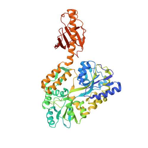
    DNA transformation is a widespread process allowing bacteria to capture free DNA by using filamentous nano-machines composed of type IV pilins. These proteins can act as DNA receptors as demonstrated by the finding that Neisseria meningitidis ComP minor pilin has intrinsic DNA-binding ability. ComP binds DNA better when it contains the DNA-uptake sequence (DUS) motif abundant in this species genome, playing a role in its trademark ability to selectively take up its own DNA. Here, we report high-resolution structures for meningococcal ComP and Neisseria subflava ComPsub, which recognize different DUS motifs. We show that they are structurally identical type IV pilins that pack readily into filament models and display a unique DD region delimited by two disulfide bonds. Functional analysis of ComPsub defines a new mode of DNA binding involving the DD region, adapted for exported DNA receptors.


  • Organizational Affiliation

    MRC Centre for Molecular Bacteriology and Infection, Imperial College London, London SW7 2AZ, UK.


Macromolecules
Find similar proteins by:  (by identity cutoff)  |  3D Structure
Entity ID: 1
MoleculeChains Sequence LengthOrganismDetailsImage
ComP490Neisseria meningitidisMutation(s): 0 
Gene Names: ComP
UniProt
Find proteins for P0AEX9 (Escherichia coli (strain K12))
Explore P0AEX9 
Go to UniProtKB:  P0AEX9
Entity Groups  
Sequence Clusters30% Identity50% Identity70% Identity90% Identity95% Identity100% Identity
UniProt GroupP0AEX9
Sequence Annotations
Expand
  • Reference Sequence
Oligosaccharides

Help

Entity ID: 2
MoleculeChains Length2D Diagram Glycosylation3D Interactions
alpha-D-glucopyranose-(1-4)-alpha-D-glucopyranose-(1-4)-alpha-D-glucopyranose
B
3N/A
Glycosylation Resources
GlyTouCan:  G96370VA
GlyCosmos:  G96370VA
GlyGen:  G96370VA
Small Molecules
Biologically Interesting Molecules (External Reference) 1 Unique
Experimental Data & Validation

Experimental Data

  • Method: X-RAY DIFFRACTION
  • Resolution: 1.43 Å
  • R-Value Free: 0.191 
  • R-Value Work: 0.157 
  • R-Value Observed: 0.158 
  • Space Group: P 21 21 21
Unit Cell:
Length ( Å )Angle ( ˚ )
a = 63.57α = 90
b = 68.44β = 90
c = 109.58γ = 90
Software Package:
Software NamePurpose
PHENIXrefinement
xia2data reduction
XSCALEdata scaling
PHASERphasing

Structure Validation

View Full Validation Report


Ligand Structure Quality Assessment 


Entry History & Funding Information

Deposition Data

  • Released Date: 2016-05-18 
  • Deposition Author(s): Berry, J.-L.

Funding OrganizationLocationGrant Number
Biotechnology and Biological Sciences Research CouncilUnited KingdomBB/J016764/

Revision History  (Full details and data files)

  • Version 1.0: 2016-05-18
    Type: Initial release
  • Version 1.1: 2016-05-25
    Changes: Database references
  • Version 1.2: 2016-06-15
    Changes: Database references
  • Version 1.3: 2017-08-30
    Changes: Author supporting evidence
  • Version 2.0: 2020-07-29
    Type: Remediation
    Reason: Carbohydrate remediation
    Changes: Advisory, Atomic model, Data collection, Derived calculations, Non-polymer description, Structure summary
  • Version 2.1: 2024-01-10
    Changes: Data collection, Database references, Refinement description, Structure summary