5K7L

Single particle cryo-EM structure of the voltage-gated K+ channel Eag1 bound to the channel inhibitor calmodulin


Experimental Data Snapshot

  • Method: ELECTRON MICROSCOPY
  • Resolution: 3.78 Å
  • Aggregation State: PARTICLE 
  • Reconstruction Method: SINGLE PARTICLE 

wwPDB Validation   3D Report Full Report


This is version 1.4 of the entry. See complete history


Literature

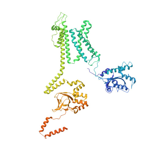
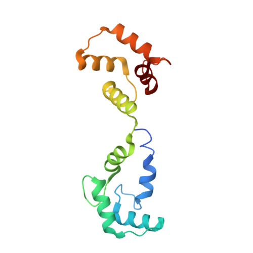
Structure of the voltage-gated K+ channel Eag1 reveals an alternative voltage sensing mechanism.

Whicher, J.R.MacKinnon, R.

(2016) Science 353: 664-669

  • DOI: https://doi.org/10.1126/science.aaf8070
  • Primary Citation of Related Structures:  
    5K7L

  • PubMed Abstract: 

    Voltage-gated potassium (K(v)) channels are gated by the movement of the transmembrane voltage sensor, which is coupled, through the helical S4-S5 linker, to the potassium pore. We determined the single-particle cryo-electron microscopy structure of mammalian K(v)10.1, or Eag1, bound to the channel inhibitor calmodulin, at 3.78 angstrom resolution. Unlike previous K(v) structures, the S4-S5 linker of Eag1 is a five-residue loop and the transmembrane segments are not domain swapped, which suggest an alternative mechanism of voltage-dependent gating. Additionally, the structure and position of the S4-S5 linker allow calmodulin to bind to the intracellular domains and to close the potassium pore, independent of voltage-sensor position. The structure reveals an alternative gating mechanism for K(v) channels and provides a template to further understand the gating properties of Eag1 and related channels.


  • Organizational Affiliation

    Laboratory of Molecular Neurobiology and Biophysics, The Rockefeller University, Howard Hughes Medical Institute, 1230 York Avenue, New York, NY 10065, USA.


Macromolecules
Find similar proteins by:  (by identity cutoff)  |  3D Structure
Entity ID: 1
MoleculeChains Sequence LengthOrganismDetailsImage
Potassium voltage-gated channel subfamily H member 1857Rattus norvegicusMutation(s): 0 
Gene Names: Kcnh1Eag
Membrane Entity: Yes 
UniProt
Find proteins for Q63472 (Rattus norvegicus)
Explore Q63472 
Go to UniProtKB:  Q63472
Entity Groups  
Sequence Clusters30% Identity50% Identity70% Identity90% Identity95% Identity100% Identity
UniProt GroupQ63472
Sequence Annotations
Expand
  • Reference Sequence
Find similar proteins by:  (by identity cutoff)  |  3D Structure
Entity ID: 2
MoleculeChains Sequence LengthOrganismDetailsImage
Calmodulin149Homo sapiensMutation(s): 0 
Gene Names: 
CALM1CALMCAMCAM1CALM2CAM2CAMBCALM3CALML2CAM3...
CALM1CALMCAMCAM1CALM2CAM2CAMBCALM3CALML2CAM3CAMCCAMIII

UniProt & NIH Common Fund Data Resources
Find proteins for P0DP23 (Homo sapiens)
Explore P0DP23 
Go to UniProtKB:  P0DP23
PHAROS:  P0DP23
GTEx:  ENSG00000198668 
Entity Groups  
Sequence Clusters30% Identity50% Identity70% Identity90% Identity95% Identity100% Identity
UniProt GroupP0DP23
Sequence Annotations
Expand
  • Reference Sequence
Small Molecules
Ligands 2 Unique
IDChains Name / Formula / InChI Key2D Diagram3D Interactions
Y01
Query on Y01

Download Ideal Coordinates CCD File 
D [auth A]CHOLESTEROL HEMISUCCINATE
C31 H50 O4
WLNARFZDISHUGS-MIXBDBMTSA-N
NAG
Query on NAG

Download Ideal Coordinates CCD File 
C [auth A]2-acetamido-2-deoxy-beta-D-glucopyranose
C8 H15 N O6
OVRNDRQMDRJTHS-FMDGEEDCSA-N
Experimental Data & Validation

Experimental Data

  • Method: ELECTRON MICROSCOPY
  • Resolution: 3.78 Å
  • Aggregation State: PARTICLE 
  • Reconstruction Method: SINGLE PARTICLE 
EM Software:
TaskSoftware PackageVersion
RECONSTRUCTIONFREALIGN
MODEL REFINEMENTREFMAC

Structure Validation

View Full Validation Report


Entry History & Funding Information

Deposition Data


Funding OrganizationLocationGrant Number
National Institutes of Health/National Institute of General Medical Sciences (NIH/NIGMS)United StatesGM43949
Howard Hughes Medical Institute (HHMI)United States--
Damon Runyon Cancer Research FoundationUnited StatesDRG-2212-15

Revision History  (Full details and data files)

  • Version 1.0: 2016-08-17
    Type: Initial release
  • Version 1.1: 2016-08-31
    Changes: Database references
  • Version 1.2: 2017-09-13
    Changes: Author supporting evidence, Data collection, Database references
  • Version 1.3: 2019-11-20
    Changes: Author supporting evidence
  • Version 1.4: 2020-07-29
    Type: Remediation
    Reason: Carbohydrate remediation
    Changes: Data collection, Derived calculations, Structure summary