Structural basis of steroid binding and oxidation by the cytochrome P450 CYP109E1 from Bacillus megaterium.
Jozwik, I.K., Kiss, F.M., Abdulmughni, A., Brill, E., Zapp, J., Pleiss, J., Bernhardt, R., Thunnissen, A.W.(2016) FEBS J 283: 4128-4148
- PubMed: 27686671
- DOI: https://doi.org/10.1111/febs.13911
- Primary Citation of Related Structures:
5L90, 5L91, 5L92, 5L94 - PubMed Abstract:
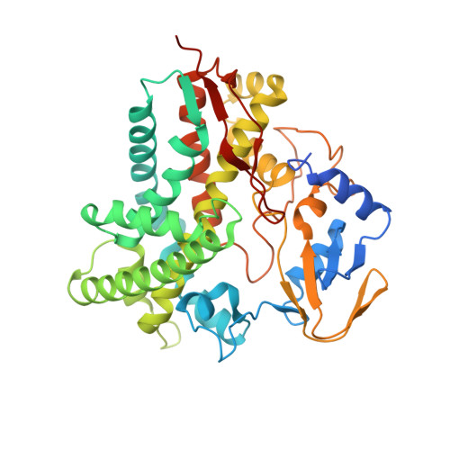
Cytochrome P450 monooxygenases (P450s) are attractive enzymes for the pharmaceutical industry, in particular, for applications in steroidal drug synthesis. Here, we report a comprehensive functional and structural characterization of CYP109E1, a novel steroid-converting cytochrome P450 enzyme identified from the genome of Bacillus megaterium DSM319. In vitro and whole-cell in vivo turnover experiments, combined with binding assays, revealed that CYP109E1 is able to hydroxylate testosterone at position 16β. Related steroids with bulky substituents at carbon C17, like corticosterone, bind to the enzyme without being converted. High-resolution X-ray structures were solved of a steroid-free form of CYP109E1 and of complexes with testosterone and corticosterone. The structural analysis revealed a highly dynamic active site at the distal side of the heme, which is wide open in the absence of steroids, can bind four ordered corticosterone molecules simultaneously, and undergoes substantial narrowing upon binding of single steroid molecules. In the crystal structures, the single bound steroids adopt unproductive binding modes coordinating the heme-iron with their C3-keto oxygen. Molecular dynamics (MD) simulations suggest that the steroids may also bind in ~180° reversed orientations with the C16 carbon and C17-substituents pointing toward the heme, leading to productive binding of testosterone explaining the observed regio- and stereoselectivity. The X-ray structures and MD simulations further identify several residues with important roles in steroid binding and conversion, which could be confirmed by site-directed mutagenesis. Taken together, our results provide unique insights into the CYP109E1 activity, substrate specificity, and regio/stereoselectivity.
Organizational Affiliation:
Laboratory of Biophysical Chemistry, Groningen Biomolecular Sciences and Biotechnology Institute, University of Groningen, The Netherlands.