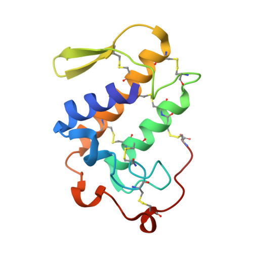
Crystal structure of a phospholipase A2 from Bothrops asper venom: Insights into a new putative "myotoxic cluster".
Salvador, G.H., Dos Santos, J.I., Lomonte, B., Fontes, M.R.(2017) Biochimie 133: 95-102
- PubMed: 28034717
- DOI: https://doi.org/10.1016/j.biochi.2016.12.015
- Primary Citation of Related Structures:
5TFV - PubMed Abstract:
Snake venoms from the Viperidae and Elapidae families often have several phospholipases A 2 (PLA 2 s), which may display different functions despite having a similar structural scaffold. These proteins are considered an important target for the development of drugs against local myotoxic damage because they are not efficiently neutralized by conventional serum therapy. PLA 2 s from these venoms are generally divided into two classes: (i) catalytic PLA 2 s (or Asp49-PLA 2 s) and (ii) non-catalytic PLA 2 -like toxins (or Lys49-PLA 2 s). In many Viperidae venoms, a subset of the basic Asp49-PLA 2 s displays some functional and structural characteristics of PLA 2 -like proteins and group within the same phylogenetic clade, but their myotoxic mechanism is still largely unknown. In the present study, we have crystallized and solved the structure of myotoxin I (MT-I), a basic myotoxic Asp49-PLA 2 isolated from Bothrops asper venom. The structure presents a dimeric conformation that is compatible with that of previous dimers found for basic myotoxic Asp49-PLA 2 s and Lys49-PLA 2 s and has been confirmed by other biophysical and bioinformatics techniques. This arrangement suggests a possible cooperative action between both monomers to exert myotoxicity via two different sites forming a putative membrane-docking site (MDoS) and a putative membrane disruption site (MDiS). This mechanism would resemble that proposed for Lys49-PLA 2 s, but the sites involved appear to be situated in a different region. Thus, as both sites are close to one another, they form a "myotoxic cluster", which is also found in two other basic myotoxic Asp49-PLA 2 s from Viperidae venoms. Such arrangement may represent a novel structural strategy for the mechanism of muscle damage exerted by the group of basic, Asp49-PLA 2 s found in viperid snake venoms.
Organizational Affiliation:
Departamento de Física e Biofísica, Instituto de Biociências, UNESP - Univ. Estadual Paulista, Botucatu, SP, Brazil.