5VB6

X-ray co-structure of nuclear receptor ROR-gammat Ligand Binding Domain with an inverse agonist and SRC2 peptide


Experimental Data Snapshot

  • Method: X-RAY DIFFRACTION
  • Resolution: 2.04 Å
  • R-Value Free: 0.207 
  • R-Value Work: 0.187 
  • R-Value Observed: 0.188 

wwPDB Validation   3D Report Full Report


Ligand Structure Quality Assessment 


This is version 1.2 of the entry. See complete history


Literature

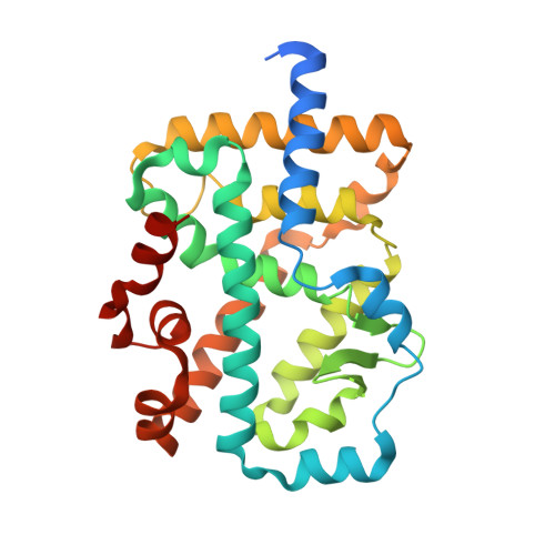
Structural studies unravel the active conformation of apo ROR gamma t nuclear receptor and a common inverse agonism of two diverse classes of ROR gamma t inhibitors.

Li, X.Anderson, M.Collin, D.Muegge, I.Wan, J.Brennan, D.Kugler, S.Terenzio, D.Kennedy, C.Lin, S.Labadia, M.E.Cook, B.Hughes, R.Farrow, N.A.

(2017) J Biol Chem 292: 11618-11630

  • DOI: https://doi.org/10.1074/jbc.M117.789024
  • Primary Citation of Related Structures:  
    5VB3, 5VB5, 5VB6, 5VB7

  • PubMed Abstract: 

    The nuclear receptor retinoid acid receptor-related orphan receptor γt (RORγt) is a master regulator of the Th17/IL-17 pathway that plays crucial roles in the pathogenesis of autoimmunity. RORγt has recently emerged as a highly promising target for treatment of a number of autoimmune diseases. Through high-throughput screening, we previously identified several classes of inverse agonists for RORγt. Here, we report the crystal structures for the ligand-binding domain of RORγt in both apo and ligand-bound states. We show that apo RORγt adopts an active conformation capable of recruiting coactivator peptides and present a detailed analysis of the structural determinants that stabilize helix 12 (H12) of RORγt in the active state in the absence of a ligand. The structures of ligand-bound RORγt reveal that binding of the inverse agonists disrupts critical interactions that stabilize H12. This destabilizing effect is supported by ab initio calculations and experimentally by a normalized crystallographic B-factor analysis. Of note, the H12 destabilization in the active state shifts the conformational equilibrium of RORγt toward an inactive state, which underlies the molecular mechanism of action for the inverse agonists reported here. Our findings highlight that nuclear receptor structure and function are dictated by a dynamic conformational equilibrium and that subtle changes in ligand structures can shift this equilibrium in opposite directions, leading to a functional switch from agonists to inverse agonists.


  • Organizational Affiliation

    Departments of Small Molecule Discovery Research, Ridgefield, Connecticut 06877-0368. Electronic address: xiang.li@boehringer-ingelheim.com.


Macromolecules
Find similar proteins by:  (by identity cutoff)  |  3D Structure
Entity ID: 1
MoleculeChains Sequence LengthOrganismDetailsImage
Nuclear receptor ROR-gamma, SRC2 chimera280Homo sapiensMutation(s): 0 
Gene Names: RORCNR1F3RORGRZRG
UniProt & NIH Common Fund Data Resources
Find proteins for P51449 (Homo sapiens)
Explore P51449 
Go to UniProtKB:  P51449
PHAROS:  P51449
GTEx:  ENSG00000143365 
Find proteins for Q15596 (Homo sapiens)
Explore Q15596 
Go to UniProtKB:  Q15596
PHAROS:  Q15596
GTEx:  ENSG00000140396 
Entity Groups  
Sequence Clusters30% Identity50% Identity70% Identity90% Identity95% Identity100% Identity
UniProt GroupsP51449Q15596
Sequence Annotations
Expand
  • Reference Sequence
Small Molecules
Ligands 2 Unique
IDChains Name / Formula / InChI Key2D Diagram3D Interactions
927
Query on 927

Download Ideal Coordinates CCD File 
C [auth A]N-{3-[(3-methylbut-2-en-1-yl){methyl[trans-4-(pyridin-4-yl)cyclohexyl]carbamoyl}amino]phenyl}benzamide
C31 H36 N4 O2
PITIPWBKTYAGSP-XBTDCMMSSA-N
NA
Query on NA

Download Ideal Coordinates CCD File 
B [auth A]SODIUM ION
Na
FKNQFGJONOIPTF-UHFFFAOYSA-N
Binding Affinity Annotations 
IDSourceBinding Affinity
927 Binding MOAD:  5VB6 Kd: 430 (nM) from 1 assay(s)
Experimental Data & Validation

Experimental Data

  • Method: X-RAY DIFFRACTION
  • Resolution: 2.04 Å
  • R-Value Free: 0.207 
  • R-Value Work: 0.187 
  • R-Value Observed: 0.188 
  • Space Group: P 41 21 2
Unit Cell:
Length ( Å )Angle ( ˚ )
a = 62.305α = 90
b = 62.305β = 90
c = 154.178γ = 90
Software Package:
Software NamePurpose
PHENIXrefinement
XDSdata reduction
Aimlessdata scaling
PHENIXphasing

Structure Validation

View Full Validation Report


Ligand Structure Quality Assessment 


Entry History 

Deposition Data

  • Released Date: 2017-06-07 
  • Deposition Author(s): Li, X.

Revision History  (Full details and data files)

  • Version 1.0: 2017-06-07
    Type: Initial release
  • Version 1.1: 2017-07-26
    Changes: Database references
  • Version 1.2: 2024-03-06
    Changes: Data collection, Database references, Derived calculations