5YD5

Crystal structure of the scFv antibody 4B08 with epitope peptide (mutation N3A)


Experimental Data Snapshot

  • Method: X-RAY DIFFRACTION
  • Resolution: 1.96 Å
  • R-Value Free: 0.258 
  • R-Value Work: 0.203 
  • R-Value Observed: 0.205 

wwPDB Validation   3D Report Full Report


This is version 1.2 of the entry. See complete history


Literature

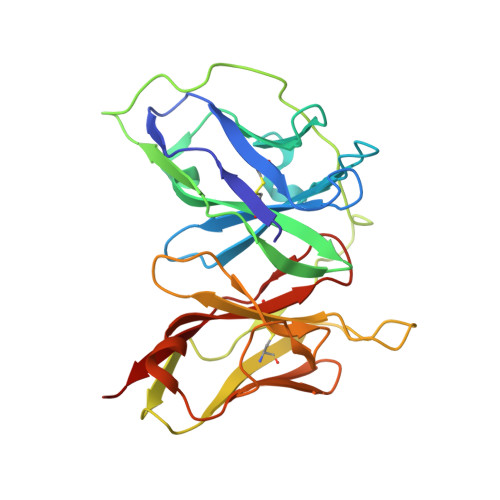
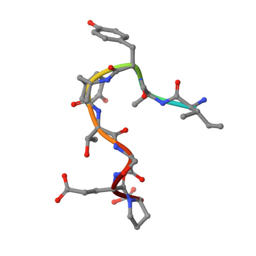
Intramolecular H-bonds govern the recognition of a flexible peptide by an antibody

Miyanabe, K.Akiba, H.Kuroda, D.Nakakido, M.Kusano-Arai, O.Iwanari, H.Hamakubo, T.Caaveiro, J.M.M.Tsumoto, K.

(2018) J Biochem 164: 65-76

  • DOI: https://doi.org/10.1093/jb/mvy032
  • Primary Citation of Related Structures:  
    5YD3, 5YD4, 5YD5

  • PubMed Abstract: 

    Molecular recognition is a fundamental event at the core of essentially every biological process. In particular, intermolecular H-bonds have been recognized as key stabilizing forces in antibody-antigen interactions resulting in exquisite specificity and high affinity. Although equally abundant, the role of intramolecular H-bonds is far less clear and not universally acknowledged. Herein, we have carried out a molecular-level study to dissect the contribution of intramolecular H-bonds in a flexible peptide for the recognition by an antibody. We show that intramolecular H-bonds may have a profound, multifaceted and favorable effect on the binding affinity by up to 2 kcal mol-1 of free energy. Collectively, our results suggest that antibodies are fine tuned to recognize transiently stabilized structures of flexible peptides in solution, for which intramolecular H-bonds play a key role.


  • Organizational Affiliation

    Department of Chemistry and Biotechnology, School of Engineering, The University of Tokyo, Bunkyo-ku, Tokyo 113-8656, Japan.


Macromolecules
Find similar proteins by:  (by identity cutoff)  |  3D Structure
Entity ID: 1
MoleculeChains Sequence LengthOrganismDetailsImage
scFv 4B08
A, C
251Mus musculusMutation(s): 0 
UniProt
Find proteins for P01630 (Mus musculus)
Explore P01630 
Go to UniProtKB:  P01630
Entity Groups  
Sequence Clusters30% Identity50% Identity70% Identity90% Identity95% Identity100% Identity
UniProt GroupP01630
Sequence Annotations
Expand
  • Reference Sequence

Find similar proteins by:  Sequence   |   3D Structure  

Entity ID: 2
MoleculeChains Sequence LengthOrganismDetailsImage
Peptide epitope (mutation N3A)
B, D
9Homo sapiensMutation(s): 0 
UniProt & NIH Common Fund Data Resources
Find proteins for P51681 (Homo sapiens)
Explore P51681 
Go to UniProtKB:  P51681
PHAROS:  P51681
GTEx:  ENSG00000160791 
Entity Groups  
UniProt GroupP51681
Sequence Annotations
Expand
  • Reference Sequence
Experimental Data & Validation

Experimental Data

  • Method: X-RAY DIFFRACTION
  • Resolution: 1.96 Å
  • R-Value Free: 0.258 
  • R-Value Work: 0.203 
  • R-Value Observed: 0.205 
  • Space Group: P 2 21 21
Unit Cell:
Length ( Å )Angle ( ˚ )
a = 61.62α = 90
b = 81.9β = 90
c = 114.22γ = 90
Software Package:
Software NamePurpose
REFMACrefinement
MOSFLMdata reduction
Aimlessdata scaling
PHASERphasing

Structure Validation

View Full Validation Report


Entry History & Funding Information

Deposition Data


Funding OrganizationLocationGrant Number
Japan Society for the Promotion of ScienceJapan25249115
Japan Society for the Promotion of ScienceJapan15K06962

Revision History  (Full details and data files)

  • Version 1.0: 2018-06-06
    Type: Initial release
  • Version 1.1: 2018-07-04
    Changes: Data collection, Database references
  • Version 1.2: 2023-11-22
    Changes: Data collection, Database references, Refinement description