


Structure of Nurr1 bound to cyclopentenone prostaglandin A2 and its mechanism of action in ameliorating dopaminergic neurodegeneration in Drosophila
Rajan, S., Toh, H.T., Lim, K.H., Yoon, H.S.To be published.
Experimental Data Snapshot
Entity ID: 1 | |||||
|---|---|---|---|---|---|
| Molecule | Chains | Sequence Length | Organism | Details | Image |
| Nuclear receptor subfamily 4 group A member 2 | 271 | Homo sapiens | Mutation(s): 0 Gene Names: NR4A2, NOT, NURR1, TINUR | 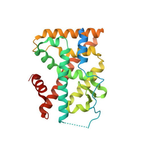 | |
UniProt & NIH Common Fund Data Resources | |||||
Find proteins for P43354 (Homo sapiens) Explore P43354 Go to UniProtKB: P43354 | |||||
PHAROS: P43354 GTEx: ENSG00000153234 | |||||
Entity Groups | |||||
| Sequence Clusters | 30% Identity50% Identity70% Identity90% Identity95% Identity100% Identity | ||||
| UniProt Group | P43354 | ||||
Sequence AnnotationsExpand | |||||
| |||||
| Ligands 2 Unique | |||||
|---|---|---|---|---|---|
| ID | Chains | Name / Formula / InChI Key | 2D Diagram | 3D Interactions | |
| 8SU (Subject of Investigation/LOI) Query on 8SU | E [auth A], H [auth B], I [auth C], J [auth D] | (~{Z})-7-[(1~{R},5~{S})-2-oxidanylidene-5-[(~{E},3~{S})-3-oxidanyloct-1-enyl]cyclopent-3-en-1-yl]hept-5-enoic acid C20 H30 O4 MYHXHCUNDDAEOZ-FOSBLDSVSA-N |  | ||
| MG Query on MG | F [auth A], G [auth B] | MAGNESIUM ION Mg JLVVSXFLKOJNIY-UHFFFAOYSA-N |  | ||
| Length ( Å ) | Angle ( ˚ ) |
|---|---|
| a = 85.95 | α = 90 |
| b = 94.08 | β = 90 |
| c = 135.65 | γ = 90 |
| Software Name | Purpose |
|---|---|
| REFMAC | refinement |
| iMOSFLM | data reduction |
| SCALA | data scaling |
| PHASER | phasing |
RCSB PDB (citation) is hosted by
RCSB PDB is a member of the

