5Z5S

Crystal structure of the PPARgamma-LBD complexed with compound 13ab


Experimental Data Snapshot

  • Method: X-RAY DIFFRACTION
  • Resolution: 1.80 Å
  • R-Value Free: 0.229 
  • R-Value Work: 0.204 
  • R-Value Observed: 0.204 

wwPDB Validation   3D Report Full Report


Ligand Structure Quality Assessment 


This is version 1.2 of the entry. See complete history


Literature

Discovery of DS-6930, a potent selective PPAR gamma modulator. Part I: Lead identification.

Shinozuka, T.Tsukada, T.Fujii, K.Tokumaru, E.Shimada, K.Onishi, Y.Matsui, Y.Wakimoto, S.Kuroha, M.Ogata, T.Araki, K.Ohsumi, J.Sawamura, R.Watanabe, N.Yamamoto, H.Fujimoto, K.Tani, Y.Mori, M.Tanaka, J.

(2018) Bioorg Med Chem 26: 5079-5098

  • DOI: https://doi.org/10.1016/j.bmc.2018.09.006
  • Primary Citation of Related Structures:  
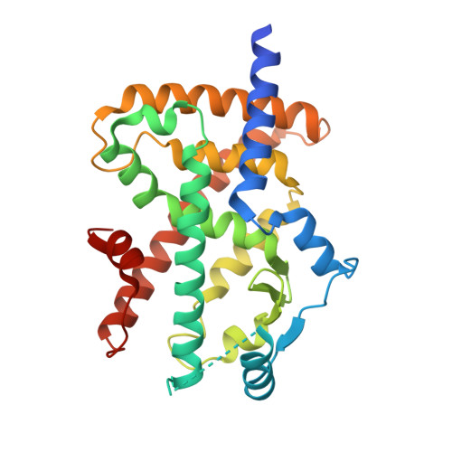
    5Z5S

  • PubMed Abstract: 

    The lead identification of a novel potent selective PPARγ agonist, DS-6930 is reported. To avoid PPARγ-related adverse effects, a partial agonist was designed to prevent the direct interaction with helix 12 of PPARγ-LBD. Because the TZD group is known to interact with helix 12, the TZD in efatutazone (CS-7017) was replaced to discover novel PPARγ intermediate partial agonist 8i. The optimization of 8i yielded 13ac with high potency in vitro. Compound 13ac exhibited robust plasma glucose lowering effects comparable to those of rosiglitazone (3 mg/kg) in Zucker diabetic fatty rats. Upon toxicological evaluation, compound 13ac (300 mg/kg) induced hemodilution to a lower extent than rosiglitazone; however, 13ac elevated liver enzyme activities. X-ray crystallography revealed no direct interaction of 13ac with helix 12, and the additional lipophilic interactions are also suggested to be related to the maximum transcriptional activity of 13ac.


  • Organizational Affiliation

    R&D Division, Daiichi Sankyo Co., Ltd., 1-2-58 Hiromachi, Shinagawa-ku, Tokyo 140-8710, Japan. Electronic address: shinozuka.tsuyoshi.s5@daiichisankyo.co.jp.


Macromolecules
Find similar proteins by:  (by identity cutoff)  |  3D Structure
Entity ID: 1
MoleculeChains Sequence LengthOrganismDetailsImage
Peroxisome proliferator-activated receptor gamma283Homo sapiensMutation(s): 0 
Gene Names: PPARGNR1C3
UniProt & NIH Common Fund Data Resources
Find proteins for P37231 (Homo sapiens)
Explore P37231 
Go to UniProtKB:  P37231
PHAROS:  P37231
GTEx:  ENSG00000132170 
Entity Groups  
Sequence Clusters30% Identity50% Identity70% Identity90% Identity95% Identity100% Identity
UniProt GroupP37231
Sequence Annotations
Expand
  • Reference Sequence

Find similar proteins by:  Sequence   |   3D Structure  

Entity ID: 2
MoleculeChains Sequence LengthOrganismDetailsImage
Peptide from Peroxisome proliferator-activated receptor gamma coactivator 1-alphaB [auth C]19Homo sapiensMutation(s): 0 
UniProt & NIH Common Fund Data Resources
Find proteins for Q9UBK2 (Homo sapiens)
Explore Q9UBK2 
Go to UniProtKB:  Q9UBK2
PHAROS:  Q9UBK2
GTEx:  ENSG00000109819 
Entity Groups  
Sequence Clusters30% Identity50% Identity70% Identity90% Identity95% Identity100% Identity
UniProt GroupQ9UBK2
Sequence Annotations
Expand
  • Reference Sequence
Small Molecules
Ligands 2 Unique
IDChains Name / Formula / InChI Key2D Diagram3D Interactions
RTE
Query on RTE

Download Ideal Coordinates CCD File 
C [auth A]3-{[6-(4-chloro-3-fluorophenoxy)-1-methyl-1H-benzimidazol-2-yl]methoxy}benzoic acid
C22 H16 Cl F N2 O4
QCDBYUFGXWPSLV-UHFFFAOYSA-N
CL
Query on CL

Download Ideal Coordinates CCD File 
D [auth A],
E [auth A]
CHLORIDE ION
Cl
VEXZGXHMUGYJMC-UHFFFAOYSA-M
Binding Affinity Annotations 
IDSourceBinding Affinity
RTE BindingDB:  5Z5S EC50: 68 (nM) from 1 assay(s)
Experimental Data & Validation

Experimental Data

  • Method: X-RAY DIFFRACTION
  • Resolution: 1.80 Å
  • R-Value Free: 0.229 
  • R-Value Work: 0.204 
  • R-Value Observed: 0.204 
  • Space Group: P 1 21 1
Unit Cell:
Length ( Å )Angle ( ˚ )
a = 43.408α = 90
b = 54.015β = 106.48
c = 66.242γ = 90
Software Package:
Software NamePurpose
CNXrefinement
HKL-2000data reduction
HKL-2000data scaling
CNXphasing

Structure Validation

View Full Validation Report


Ligand Structure Quality Assessment 


Entry History 

Deposition Data

Revision History  (Full details and data files)

  • Version 1.0: 2018-10-17
    Type: Initial release
  • Version 1.1: 2018-10-24
    Changes: Data collection, Database references
  • Version 1.2: 2023-11-22
    Changes: Data collection, Database references, Refinement description