


Crystal structure of LdBPK_091320.1 with with inhibitor bound
Lin, Y.H., Dong, A., Loppnau, P., Bountra, C., Arrowsmith, C.H., Edwards, A.M., Hui, R., Structural Genomics Consortium (SGC)To be published.
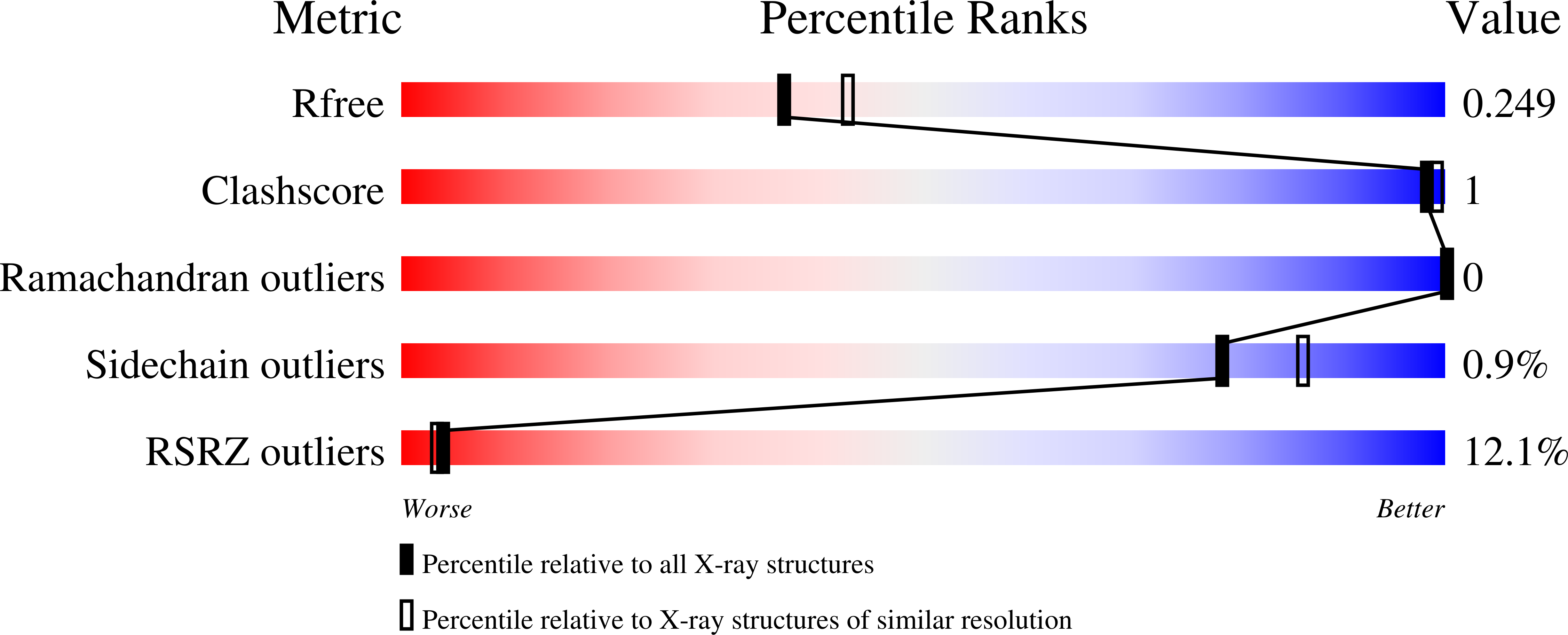
Experimental Data Snapshot
Entity ID: 1 | |||||
|---|---|---|---|---|---|
| Molecule | Chains | Sequence Length | Organism | Details | Image |
| Uncharacterized protein | 133 | Leishmania donovani BPK282A1 | Mutation(s): 0 Gene Names: LDBPK_091320 | 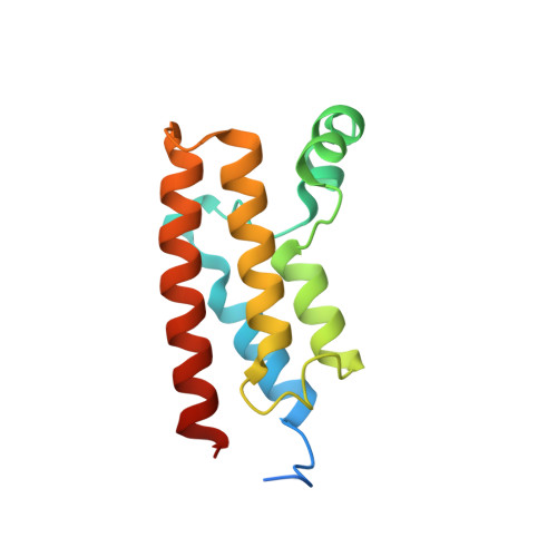 | |
UniProt | |||||
Find proteins for A0A3Q8I8I6 (Leishmania donovani) Explore A0A3Q8I8I6 Go to UniProtKB: A0A3Q8I8I6 | |||||
Entity Groups | |||||
| Sequence Clusters | 30% Identity50% Identity70% Identity90% Identity95% Identity100% Identity | ||||
| UniProt Group | A0A3Q8I8I6 | ||||
Sequence AnnotationsExpand | |||||
| |||||
| Ligands 2 Unique | |||||
|---|---|---|---|---|---|
| ID | Chains | Name / Formula / InChI Key | 2D Diagram | 3D Interactions | |
| 2LO Query on 2LO | E [auth A], G [auth B], I [auth C], K [auth D] | 2-[2-(3-chloro-4-methoxyphenyl)ethyl]-5-(3,5-dimethyl-1,2-oxazol-4-yl)-1-[(2S)-2-(morpholin-4-yl)propyl]-1H-benzimidazole C28 H33 Cl N4 O3 GEPYBHCJBORHCE-SFHVURJKSA-N |  | ||
| UNX Query on UNX | F [auth A], H [auth B], J [auth C] | UNKNOWN ATOM OR ION X |  | ||
| Length ( Å ) | Angle ( ˚ ) |
|---|---|
| a = 82.615 | α = 90 |
| b = 82.615 | β = 90 |
| c = 177.235 | γ = 90 |
| Software Name | Purpose |
|---|---|
| BUSTER | refinement |
| SCALEPACK | data scaling |
| PDB_EXTRACT | data extraction |
| HKL-3000 | data reduction |
| PHASER | phasing |
RCSB PDB (citation) is hosted by
RCSB PDB is a member of the

