


Crystallographic selection of adenosine analogs that fit the mold of the active site of human GRP78 and beyond
Antoshchenko, T., Chen, Y., Park, H.To be published.
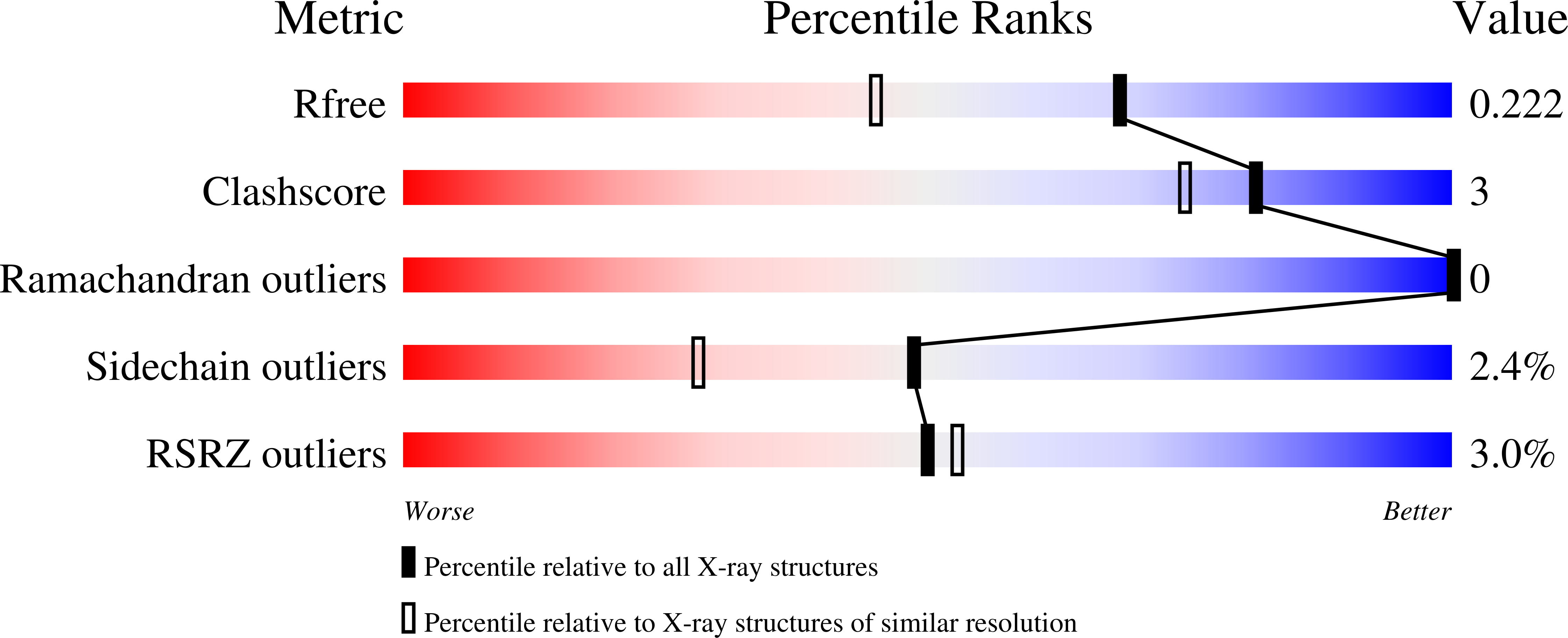
Experimental Data Snapshot
Entity ID: 1 | |||||
|---|---|---|---|---|---|
| Molecule | Chains | Sequence Length | Organism | Details | Image |
| Endoplasmic reticulum chaperone BiP | 382 | Homo sapiens | Mutation(s): 0 Gene Names: HSPA5, GRP78 EC: 3.6.4.10 | 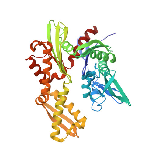 | |
UniProt & NIH Common Fund Data Resources | |||||
Find proteins for P11021 (Homo sapiens) Explore P11021 Go to UniProtKB: P11021 | |||||
PHAROS: P11021 GTEx: ENSG00000044574 | |||||
Entity Groups | |||||
| Sequence Clusters | 30% Identity50% Identity70% Identity90% Identity95% Identity100% Identity | ||||
| UniProt Group | P11021 | ||||
Sequence AnnotationsExpand | |||||
| |||||
| Ligands 2 Unique | |||||
|---|---|---|---|---|---|
| ID | Chains | Name / Formula / InChI Key | 2D Diagram | 3D Interactions | |
| 3FD Query on 3FD | C [auth A], E [auth B] | 4-[[(2R,3S,4R,5R)-5-[6-amino-8-[(3,4-dichlorophenyl)methylamino]purin-9-yl]-3,4-dihydroxy-oxolan-2-yl]methoxymethyl]benzonitrile C25 H23 Cl2 N7 O4 ZXGGCBQORXDVTE-UMCMBGNQSA-N |  | ||
| MG Query on MG | D [auth A], F [auth B] | MAGNESIUM ION Mg JLVVSXFLKOJNIY-UHFFFAOYSA-N |  | ||
| Length ( Å ) | Angle ( ˚ ) |
|---|---|
| a = 55.28 | α = 90 |
| b = 74.92 | β = 98.52 |
| c = 86.2 | γ = 90 |
| Software Name | Purpose |
|---|---|
| REFMAC | refinement |
| PDB_EXTRACT | data extraction |
| XDS | data reduction |
| SCALA | data scaling |
| MOLREP | phasing |
RCSB PDB (citation) is hosted by
RCSB PDB is a member of the

