6GXJ

X-ray structure of DiRu-1-encapsulated Apoferritin


Experimental Data Snapshot

  • Method: X-RAY DIFFRACTION
  • Resolution: 1.43 Å
  • R-Value Free: 0.191 
  • R-Value Work: 0.174 
  • R-Value Observed: 0.174 

wwPDB Validation   3D Report Full Report


This is version 1.2 of the entry. See complete history


Literature

Encapsulation of the Dinuclear Trithiolato-Bridged Arene Ruthenium Complex Diruthenium-1 in an Apoferritin Nanocage: Structure and Cytotoxicity.

Petruk, G.Monti, D.M.Ferraro, G.Pica, A.D'Elia, L.Pane, F.Amoresano, A.Furrer, J.Kowalski, K.Merlino, A.

(2019) ChemMedChem 14: 594-602

  • DOI: https://doi.org/10.1002/cmdc.201800805
  • Primary Citation of Related Structures:  
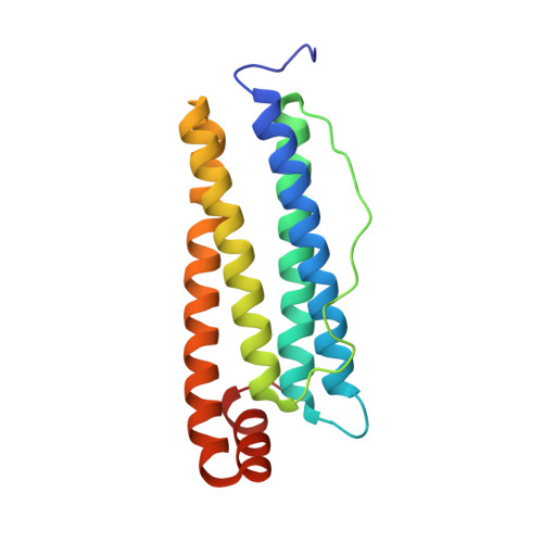
    6GXJ

  • PubMed Abstract: 

    The effects of encapsulating the cytotoxic dinuclear trithiolato-bridged arene ruthenium complex [(η 6 -p-MeC 6 H 4 iPr) 2 Ru 2 2 -S-p-C 6 H 4 tBu) 3 ]Cl (DiRu-1) within the apoferritin (AFt) nanocage were investigated. The DiRu-1-AFt nanocarrier was characterized by UV/Vis spectroscopy, ICP-MS, CD and X-ray crystallography. In contrast to previously reported Au- and Pt-based drug-loaded AFt carriers, we found no evidence of direct interactions between DiRu-1 and AFt. DiRu-1-AFt is cytotoxic toward immortalized murine BALB/c-3T3 fibroblasts transformed with SV40 virus (SVT2) and human epidermoid carcinoma A431 malignant cells, and exhibits moderate selectivity for these cancer cells over normal BALB/c-3T3 cells. DiRu-1-AFt triggers the production of reactive oxygen species, depolarization of mitochondrial membrane potential, and induces cell death via p53-mediated apoptosis. Comparison between our data and previous results suggests that the presence of specific interactions between a metal-based drug and AFt within the protein cage is not essential for drug encapsulation.


  • Organizational Affiliation

    Department of Chemical Sciences, University of Naples Federico II, Complesso Universitario di Monte Sant'Angelo, via Cinthia 21, 80126, Naples, Italy.


Macromolecules
Find similar proteins by:  (by identity cutoff)  |  3D Structure
Entity ID: 1
MoleculeChains Sequence LengthOrganismDetailsImage
Ferritin light chain174Equus caballusMutation(s): 0 
UniProt
Find proteins for P02791 (Equus caballus)
Explore P02791 
Go to UniProtKB:  P02791
Entity Groups  
Sequence Clusters30% Identity50% Identity70% Identity90% Identity95% Identity100% Identity
UniProt GroupP02791
Sequence Annotations
Expand
  • Reference Sequence
Small Molecules
Ligands 4 Unique
IDChains Name / Formula / InChI Key2D Diagram3D Interactions
CD
Query on CD

Download Ideal Coordinates CCD File 
B [auth A]
C [auth A]
D [auth A]
E [auth A]
F [auth A]
B [auth A],
C [auth A],
D [auth A],
E [auth A],
F [auth A],
G [auth A],
H [auth A],
I [auth A],
J [auth A],
M [auth A]
CADMIUM ION
Cd
WLZRMCYVCSSEQC-UHFFFAOYSA-N
SO4
Query on SO4

Download Ideal Coordinates CCD File 
N [auth A]SULFATE ION
O4 S
QAOWNCQODCNURD-UHFFFAOYSA-L
GOL
Query on GOL

Download Ideal Coordinates CCD File 
O [auth A],
P [auth A]
GLYCEROL
C3 H8 O3
PEDCQBHIVMGVHV-UHFFFAOYSA-N
CL
Query on CL

Download Ideal Coordinates CCD File 
K [auth A],
L [auth A]
CHLORIDE ION
Cl
VEXZGXHMUGYJMC-UHFFFAOYSA-M
Experimental Data & Validation

Experimental Data

  • Method: X-RAY DIFFRACTION
  • Resolution: 1.43 Å
  • R-Value Free: 0.191 
  • R-Value Work: 0.174 
  • R-Value Observed: 0.174 
  • Space Group: F 4 3 2
Unit Cell:
Length ( Å )Angle ( ˚ )
a = 180.404α = 90
b = 180.404β = 90
c = 180.404γ = 90
Software Package:
Software NamePurpose
BUSTERrefinement
XDSdata reduction
XSCALEdata scaling
BUSTERphasing

Structure Validation

View Full Validation Report


Entry History 

Deposition Data

Revision History  (Full details and data files)

  • Version 1.0: 2019-02-06
    Type: Initial release
  • Version 1.1: 2019-03-13
    Changes: Data collection, Database references
  • Version 1.2: 2024-01-17
    Changes: Data collection, Database references, Derived calculations, Refinement description