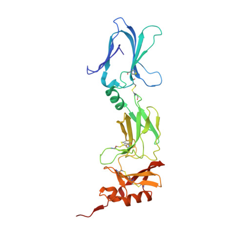
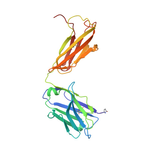
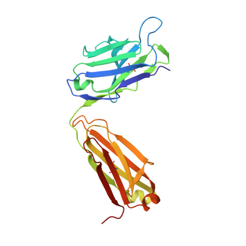
6HGA

Crystal Structure of the human IL-17RC D2-D3-D4 domains in complex with an anti-APP tag Fab


Experimental Data Snapshot

  • Method: X-RAY DIFFRACTION
  • Resolution: 2.60 Å
  • R-Value Free: 0.232 
  • R-Value Work: 0.205 
  • R-Value Observed: 0.206 

wwPDB Validation   3D Report Full Report


Ligand Structure Quality Assessment 


This is version 1.2 of the entry. See complete history


Literature

Structural Analysis Reveals that the Cytokine IL-17F Forms a Homodimeric Complex with Receptor IL-17RC to Drive IL-17RA-Independent Signaling.

Goepfert, A.Lehmann, S.Blank, J.Kolbinger, F.Rondeau, J.M.

(2020) Immunity 52: 499-512.e5

  • DOI: https://doi.org/10.1016/j.immuni.2020.02.004
  • Primary Citation of Related Structures:  
    6HG4, 6HG9, 6HGA, 6HGO, 6HGU

  • PubMed Abstract: 

    Interleukin-17A (IL-17A), IL-17F, and IL-17A/F heterodimers are key cytokines of the innate and adaptive immune response. Dysregulation of the IL-17 pathway contributes to immune pathology, and it is therefore important to elucidate the molecular mechanisms that govern IL-17 recognition and signaling. The receptor IL-17RC is thought to act in concert with IL-17RA to transduce IL-17A-, IL-17F-, and IL-17A/F-mediated signals. We report the crystal structure of the extracellular domain of human IL-17RC in complex with IL-17F. In contrast to the expected model, we found that IL-17RC formed a symmetrical 2:1 complex with IL-17F, thus competing with IL-17RA for cytokine binding. Using biophysical techniques, we showed that IL-17A and IL-17A/F also form 2:1 complexes with IL-17RC, suggesting the possibility of IL-17RA-independent IL-17 signaling pathways. The crystal structure of the IL-17RC:IL-17F complex provides a structural basis for IL-17F signaling through IL-17RC, with potential therapeutic applications for respiratory allergy and inflammatory bowel diseases.


  • Organizational Affiliation

    Novartis Institutes for BioMedical Research, Novartis Pharma AG, 4002 Basel, Switzerland.


Macromolecules
Find similar proteins by:  (by identity cutoff)  |  3D Structure
Entity ID: 1
MoleculeChains Sequence LengthOrganismDetailsImage
Interleukin-17 receptor CA [auth B]264Homo sapiensMutation(s): 5 
Gene Names: IL17RCUNQ6118/PRO20040/PRO38901
Membrane Entity: Yes 
UniProt & NIH Common Fund Data Resources
Find proteins for Q8NAC3 (Homo sapiens)
Explore Q8NAC3 
Go to UniProtKB:  Q8NAC3
PHAROS:  Q8NAC3
GTEx:  ENSG00000163702 
Entity Groups  
Sequence Clusters30% Identity50% Identity70% Identity90% Identity95% Identity100% Identity
UniProt GroupQ8NAC3
Sequence Annotations
Expand
  • Reference Sequence
Find similar proteins by:  (by identity cutoff)  |  3D Structure
Entity ID: 2
MoleculeChains Sequence LengthOrganismDetailsImage
anti-APP-tag Fab heavy-chainB [auth H]222Mus musculusMutation(s): 0 
Entity Groups  
Sequence Clusters30% Identity50% Identity70% Identity90% Identity95% Identity100% Identity
Sequence Annotations
Expand
  • Reference Sequence
Find similar proteins by:  (by identity cutoff)  |  3D Structure
Entity ID: 3
MoleculeChains Sequence LengthOrganismDetailsImage
anti-APP-tag Fab light-chainC [auth L]219Mus musculusMutation(s): 0 
Entity Groups  
Sequence Clusters30% Identity50% Identity70% Identity90% Identity95% Identity100% Identity
Sequence Annotations
Expand
  • Reference Sequence
Small Molecules
Ligands 1 Unique
IDChains Name / Formula / InChI Key2D Diagram3D Interactions
NAG
Query on NAG

Download Ideal Coordinates CCD File 
D [auth B]2-acetamido-2-deoxy-beta-D-glucopyranose
C8 H15 N O6
OVRNDRQMDRJTHS-FMDGEEDCSA-N
Modified Residues  1 Unique
IDChains TypeFormula2D DiagramParent
PCA
Query on PCA
B [auth H]L-PEPTIDE LINKINGC5 H7 N O3GLN
Experimental Data & Validation

Experimental Data

  • Method: X-RAY DIFFRACTION
  • Resolution: 2.60 Å
  • R-Value Free: 0.232 
  • R-Value Work: 0.205 
  • R-Value Observed: 0.206 
  • Space Group: P 62 2 2
Unit Cell:
Length ( Å )Angle ( ˚ )
a = 250.91α = 90
b = 250.91β = 90
c = 78.61γ = 120
Software Package:
Software NamePurpose
XDSdata reduction
XSCALEdata scaling
PHASERphasing
BUSTERrefinement
PDB_EXTRACTdata extraction

Structure Validation

View Full Validation Report


Ligand Structure Quality Assessment 


Entry History 

Deposition Data

Revision History  (Full details and data files)

  • Version 1.0: 2020-03-18
    Type: Initial release
  • Version 1.1: 2020-04-01
    Changes: Database references
  • Version 1.2: 2020-07-29
    Type: Remediation
    Reason: Carbohydrate remediation
    Changes: Data collection, Derived calculations, Structure summary