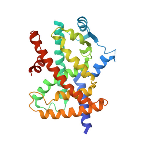
6JF0

Covalent labeling of hPPARg-LBD by turn-on fluorescent probe mediated by conjugate addition and cyclization


Experimental Data Snapshot

  • Method: X-RAY DIFFRACTION
  • Resolution: 3.40 Å
  • R-Value Free: 0.296 
  • R-Value Work: 0.267 
  • R-Value Observed: 0.268 

wwPDB Validation   3D Report Full Report


Ligand Structure Quality Assessment 


This is version 1.2 of the entry. See complete history


Literature

Cyclization Reaction-Based Turn-on Probe for Covalent Labeling of Target Proteins.

Kojima, H.Fujita, Y.Takeuchi, R.Ikebe, Y.Ohashi, N.Yamamoto, K.Itoh, T.

(2020) Cell Chem Biol 27: 334

  • DOI: https://doi.org/10.1016/j.chembiol.2020.01.006
  • Primary Citation of Related Structures:  
    6JEY, 6JEZ, 6JF0

  • PubMed Abstract: 

    Fluorescent molecules have contributed to basic biological research but there are currently only a limited number of probes available for the detection of non-enzymatic proteins. Here, we report turn-on fluorescent probes mediated by conjugate addition and cyclization (TCC probes). These probes react with multiple amino acids and exhibit a 36-fold greater emission intensity after reaction. We analyzed the reactions between TCC probes and nuclear receptors by electrospray ionization mass spectrometry, X-ray crystallography, spectrofluorometry, and fluorescence microscopy. In vitro analysis showed that probes consisting of a protein ligand and TCC could label vitamin D receptor and peroxisome proliferator-activated receptor γ. Moreover, we demonstrated that not only a ligand unit but also a peptide unit can label the target protein in a complex mixture.


  • Organizational Affiliation

    Showa Pharmaceutical University, 3-3165 Higashi-Tamagawagakuen, Machida, Tokyo 194-8543, Japan.


Macromolecules
Find similar proteins by:  (by identity cutoff)  |  3D Structure
Entity ID: 1
MoleculeChains Sequence LengthOrganismDetailsImage
Peroxisome proliferator-activated receptor gamma
A, B
276Homo sapiensMutation(s): 0 
Gene Names: PPARGNR1C3
UniProt & NIH Common Fund Data Resources
Find proteins for P37231 (Homo sapiens)
Explore P37231 
Go to UniProtKB:  P37231
PHAROS:  P37231
GTEx:  ENSG00000132170 
Entity Groups  
Sequence Clusters30% Identity50% Identity70% Identity90% Identity95% Identity100% Identity
UniProt GroupP37231
Sequence Annotations
Expand
  • Reference Sequence
Small Molecules
Ligands 2 Unique
IDChains Name / Formula / InChI Key2D Diagram3D Interactions
EB6
Query on EB6

Download Ideal Coordinates CCD File 
C [auth A]methyl (2~{S})-3-[4-[3-(4-methoxy-2-oxidanyl-phenyl)prop-2-ynoyloxy]phenyl]-2-[[2-(phenylcarbonyl)phenyl]amino]propanoate
C33 H27 N O7
CEHNFAIQCQNZKE-LJAQVGFWSA-N
EBF
Query on EBF

Download Ideal Coordinates CCD File 
D [auth B]7-methoxychromen-2-one
C10 H8 O3
LIIALPBMIOVAHH-UHFFFAOYSA-N
Experimental Data & Validation

Experimental Data

  • Method: X-RAY DIFFRACTION
  • Resolution: 3.40 Å
  • R-Value Free: 0.296 
  • R-Value Work: 0.267 
  • R-Value Observed: 0.268 
  • Space Group: C 1 2 1
Unit Cell:
Length ( Å )Angle ( ˚ )
a = 92.12α = 90
b = 61.23β = 102.26
c = 117.37γ = 90
Software Package:
Software NamePurpose
REFMACrefinement
iMOSFLMdata reduction
Aimlessdata scaling
REFMACphasing

Structure Validation

View Full Validation Report


Ligand Structure Quality Assessment 


Entry History 

Deposition Data

Revision History  (Full details and data files)

  • Version 1.0: 2020-02-12
    Type: Initial release
  • Version 1.1: 2020-04-08
    Changes: Database references
  • Version 1.2: 2023-11-22
    Changes: Data collection, Database references, Refinement description