6NKT

Ternary complex crystal structure of K289M variant of DNA polymerase Beta with beta-gamma difluoro analogue of dGTP


Experimental Data Snapshot

  • Method: X-RAY DIFFRACTION
  • Resolution: 2.60 Å
  • R-Value Free: 0.303 
  • R-Value Work: 0.199 
  • R-Value Observed: 0.209 

wwPDB Validation   3D Report Full Report


Ligand Structure Quality Assessment 


This is version 1.2 of the entry. See complete history


Literature

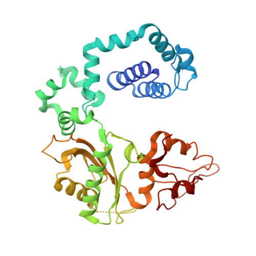
Revealing an Internal Stabilization Deficiency in the DNA Polymerase beta K289M Cancer Variant through the Combined Use of Chemical Biology and X-ray Crystallography.

Batra, V.K.Alnajjar, K.S.Sweasy, J.B.McKenna, C.E.Goodman, M.F.Wilson, S.H.

(2020) Biochemistry 59: 955-963

  • DOI: https://doi.org/10.1021/acs.biochem.9b01072
  • Primary Citation of Related Structures:  
    6NKR, 6NKS, 6NKT, 6NKU, 6NKV, 6NKW, 6NKX, 6NKY, 6NKZ, 6NL0

  • PubMed Abstract: 

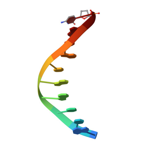
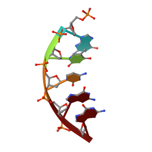
    The human DNA polymerase (pol) β cancer variant K289M has altered polymerase activity in vitro , and the structure of wild-type pol β reveals that the K289 side chain contributes to a network of stabilizing interactions in a C-terminal region of the enzyme distal to the active site. Here, we probed the capacity of the K289M variant to tolerate strain introduced within the C-terminal region and active site. Strain was imposed by making use of a dGTP analogue containing a CF 2 group substitution for the β-γ bridging oxygen atom. The ternary complex structure of the K289M variant displays an alteration in the C-terminal region, whereas the structure of wild-type pol β is not altered in the presence of the dGTP CF 2 analogue. The alteration in the K289M variant impacts the active site, because the enzyme in the ternary complex fails to adopt the normal open to closed conformational change and assembly of the catalytically competent active site. These results reveal the importance of the K289-mediated stabilizing network in the C-terminal region of pol β and suggest an explanation for why the K289M cancer variant is deficient in polymerase activity even though the position 289 side chain is distal to the active site.


  • Organizational Affiliation

    Genome Integrity and Structural Biology Laboratory, National Institute of Environmental Health Sciences, National Institutes of Health, 111 T. W. Alexander Drive, Research Triangle Park, North Carolina 27709, United States.


Macromolecules

Find similar proteins by:  (by identity cutoff)  |  3D Structure
Entity ID: 1
MoleculeChains Sequence LengthOrganismDetailsImage
DNA polymerase beta335Homo sapiensMutation(s): 1 
Gene Names: POLB
EC: 2.7.7.7 (PDB Primary Data), 4.2.99 (PDB Primary Data)
UniProt & NIH Common Fund Data Resources
Find proteins for P06746 (Homo sapiens)
Explore P06746 
Go to UniProtKB:  P06746
PHAROS:  P06746
GTEx:  ENSG00000070501 
Entity Groups  
Sequence Clusters30% Identity50% Identity70% Identity90% Identity95% Identity100% Identity
UniProt GroupP06746
Sequence Annotations
Expand
  • Reference Sequence

Find similar nucleic acids by:  Sequence   |   3D Structure  

Entity ID: 2
MoleculeChains LengthOrganismImage
DNA (5'-D(*CP*CP*GP*AP*CP*CP*GP*CP*GP*CP*AP*TP*CP*AP*GP*C)-3')B [auth T]16Homo sapiens
Sequence Annotations
Expand
  • Reference Sequence

Find similar nucleic acids by:  Sequence   |   3D Structure  

Entity ID: 3
MoleculeChains LengthOrganismImage
DNA (5'-D(*GP*CP*TP*GP*AP*TP*GP*CP*GP*(DOC))-3')C [auth P]10Homo sapiens
Sequence Annotations
Expand
  • Reference Sequence

Find similar nucleic acids by:  Sequence   |   3D Structure  

Entity ID: 4
MoleculeChains LengthOrganismImage
DNA (5'-D(P*GP*TP*CP*GP*G)-3')5Homo sapiens
Sequence Annotations
Expand
  • Reference Sequence
Small Molecules
Ligands 2 Unique
IDChains Name / Formula / InChI Key2D Diagram3D Interactions
GFF
Query on GFF

Download Ideal Coordinates CCD File 
G [auth T]2'-DEOXY-5'-O-[({[DIFLUORO(PHOSPHONO)METHYL](HYDROXY)PHOSPHORYL}OXY)(HYDROXY)PHOSPHORYL]GUANOSINE
C11 H16 F2 N5 O12 P3
BLHFKUNEUAHYSE-KVQBGUIXSA-N
NA
Query on NA

Download Ideal Coordinates CCD File 
E [auth A],
F [auth A]
SODIUM ION
Na
FKNQFGJONOIPTF-UHFFFAOYSA-N
Experimental Data & Validation

Experimental Data

  • Method: X-RAY DIFFRACTION
  • Resolution: 2.60 Å
  • R-Value Free: 0.303 
  • R-Value Work: 0.199 
  • R-Value Observed: 0.209 
  • Space Group: P 1 21 1
Unit Cell:
Length ( Å )Angle ( ˚ )
a = 55.33α = 90
b = 79.669β = 106.36
c = 54.673γ = 90
Software Package:
Software NamePurpose
HKL-2000data reduction
HKL-2000data scaling
PHENIXrefinement
PDB_EXTRACTdata extraction
PHENIXphasing

Structure Validation

View Full Validation Report


Ligand Structure Quality Assessment 


Entry History 

Deposition Data

Revision History  (Full details and data files)

  • Version 1.0: 2020-01-08
    Type: Initial release
  • Version 1.1: 2020-07-22
    Changes: Database references
  • Version 1.2: 2024-03-13
    Changes: Advisory, Data collection, Database references, Derived calculations